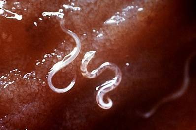
Ancylostoma caninum is a round worm that belongs to the phylum Nematoda and is found mainly distributed in the tropical and subtropical zones of the planet. This is so because those regions are the ones that meet the necessary environmental conditions for their eggs to develop effectively..
As with a large number of nematodes, Ancylostoma caninum it requires a host to develop, the dog being the perfect space for it. Inside this it fixes in the intestine and feeds on its blood.
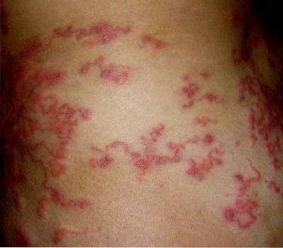
This parasite is responsible for causing intestinal disorders in dogs and can occasionally infect humans, generating serious skin lesions.
Article index
Ancylostoma caninum it is an organism that belongs to the Eukarya kingdom. As such, your cells have a structure within the cytoplasm known as the cell nucleus. Within this are the chromosomes, which are made up of DNA.
Likewise, this is a parasitic life animal, which implies that it cannot live freely, but necessarily associated with a host..
In addition, it is a heterotrophic organism because it does not have the ability to synthesize its own nutrients, so it must feed on other living beings or substances made by others. In this sense, Ancylostoma caninum, it is hematophagous, as it feeds on the blood of its host.
This parasite reproduces sexually, they are oviparous and have an indirect development. When the eggs hatch, larvae emerge from them that must undergo certain transformations or molts until they reach adulthood and be able to reproduce..
During its embryonic development, the three germ layers can be seen: ectoderm, mesoderm and endoderm, from which the tissues that will make up adult individuals originate. In the same way, this parasite is a pseudocoelomed organism, which implies that it has an internal cavity, whose origin is not mesodermal..
The taxonomic classification of Ancyllostoma caninum is the next:
-Domain: Eukarya
-Animalia Kingdom
-Phylum: Nematoda
-Class: Secernentea
-Order: Strongylida
-Family: Anclomuiosida
-Gender: Ancylostoma
-Species: Ancylostoma caninum
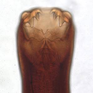
Nematodes are worms that are cylindrical in shape. Its body is covered by a resistant and protective white cuticle. They have a buccal capsule, which contains about three pairs of teeth.
They have sexual dimorphism, due to which females and males present morphological differences. The female's tail ends straight, while the male's has a structure known as a copulating bag..
As with most nematodes, females are larger than males. They measure approximately 16 mm, while males are only 10 mm.
The biological cycle of Ancylostoma caninum it is of the direct type. This means that the infective form of the larvae develops in the environment..
This parasite does not require a vector, but it does require a definitive host. In this case, the host is the dog.
The eggs are released to the outside through the feces. Depending on the humidity and temperature conditions of the soil, these can hatch, thus freeing the larvae that are covered by a protective cuticle. This occurs between day 2 and 9 after the eggs have been released..
Later, in an approximate period of about 5 days, the larvae undergo two transformations and pass to the L3 larval stage, that is, the infective form. It is understood that the larvae do not remain in the feces, but instead move to the ground, where they can remain for several days, even weeks, waiting for a host to infect. Of course, as long as the humidity and temperature conditions are ideal (humid and cool).
Once a host appears, specifically a dog, the larvae are able to enter its body and infect it.
The larvae have the ability to enter the host through the skin, mainly through the space between the pads of the legs, which are in constant contact with the ground. They penetrate the hair follicles and join the dermis (the deepest layer of the skin). Then they move through it until they enter an artery or vein, thus attaching themselves to the bloodstream..
Through the blood they are transported to the lungs, where they leave the blood and pass to the alveoli. They then ascend through the respiratory tract (bronchioles, trachea bronchi), until they reach the pharynx to be swallowed and thus pass to the digestive system.
Through the esophagus, the larvae reach the stomach and then the intestine. Here they undergo another transformation, thus becoming adult organisms, already capable of producing eggs. The adult parasite attaches to the intestinal wall through the mouth capsule. There it feeds on the blood of its host.
Ancylostoma caninum It is the parasite responsible for an infection in dogs and cats that mainly affects their digestive tract. In humans, it causes a pathology called larva migrans, which is caused mainly by the migration and displacement of the larvae through the tissues of the individual..
Humans are mainly infected by walking barefoot in places where larvae of this parasite can be found. The contagion from an animal to humans by direct contact between the two has not yet been proven..
The symptoms that dogs that are infected by this parasite present are the following:
- Anemia, caused by loss of blood from the intestine.
- Clotting disorders, caused by the secretion of anticoagulants by the parasite.
- Constant liquid stools, often showing traces of blood.
- Weakness and apathy.
- Dehydration.
- Dark colored stools from blood loss.
- Pale mucous membranes, which is also caused by loss of blood at the intestinal level.
The symptoms that human beings present are related to the damage caused by the larvae as they move through the tissues, mainly through the skin. The signs and symptoms are:
- Lesions on the skin, which are red lines and represent the displacement of the larvae through the tissues of the epidermis.
- Excruciating itching in the aforementioned lesions.
- Bacterial infections in the initial lesions.
In general, the larvae die in a short time, so there is no opportunity for them to affect internal organs of the individual beyond the skin.
Taking into account that Ancylostoma caninum it is a parasite, the infections it causes are treated with anthelmintic drugs. Among the drugs most used to treat the disease are benzimidazoles, emodepside and endectocides..
As with most parasites, the most commonly used drugs are albendazole and fenbendazole. These drugs act by causing a degeneration and destruction of certain organelles of the parasites, which results in the eventual death of the adult parasites and the larvae produced by them..


Yet No Comments