
The camel spiders or solífugos are a group of arachnids that are characterized by having very well developed chelicerae (typical appendages of some arthropods) that are very useful in their feeding process. They were first studied and described by the Swedish zoologist Carl Jakob Sundevall around 1833.
They differ from other arachnids in that they do not have venom glands in their chelicerae and their pedipalps are similar to legs, but they end in a suction cup-like structure that allows them to adhere to their prey..
Article index
Solifuges are a group of animals that belong to the so-called multicellular eukaryotic organisms. This means that in your cells, the genetic material is enclosed within the nucleus, forming chromosomes..
Likewise, solifuges are made up of various types of cells, each of which specializes in a specific function. This occurs since embryonic development, thanks to the fact that this organism presents the three germ layers: ectoderm, mesoderm and endoderm..
Continuing with embryonic development, solifuges are considered deuterostomized, because the same embryonic structure (blastopore) simultaneously gives rise to both the mouth and the anus..
Taking nutrition into account, solifuges are heterotrophs, since they are unable to synthesize their nutrients. Therefore, they must feed on other living beings or substances made by others. In this sense, these animals are carnivores and very good predators..
Anatomically, solifuges are dioecious. This implies that there are individuals with female reproductive organs and individuals with male reproductive organs..
As with many eukaryotic organisms, solifuges have bilateral symmetry. This is because they are made up of two exactly the same halves.
Domain: Eukarya
Animalia Kingdom
Phylum: Arthropoda
Subphylum: Chellicerata
Class: Arachnida
Order: Solifugae.
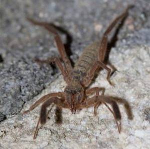
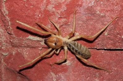
Camel spiders are characterized by having an elongated-looking body, being large (they reach up to 7 cm in length) and having a voluminous abdomen. In appearance they are similar to spiders, although they differ greatly from these.
As with the rest of the arachnids, the body of the solifuge is divided into two parts: prosoma and opistosome. The prosome is the anterior portion, while the opistosome is the posterior.
These animals are characterized by presenting very prominent and developed chelicerae.
The prosome of this type of arachnid is small. This is covered by a kind of exoskeleton or shell, whose segments are not fused..
This shell is made up of three plates, the most anterior being the propeltidium, immediately after this is the mesopeltidium and then the postpeltidium. The propeltidium presents in its anterior border the organs of sight of the animal.
The ventral surface of the prosoma is almost entirely occupied by the first joints (coxa) of the locomotor appendages of the animal..
From the prosoma emerge all the articulated appendages that are represented by the chelicerae, the pedipalps and the legs..
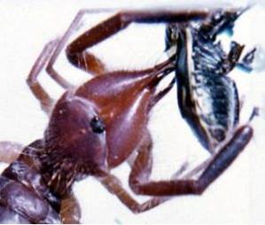
They are one of the characteristic elements of the animals of this order. They are highly developed and robust.
They are made up of two pieces. In addition, at their terminal end they have structures known as teeth. These are classified into anterior, intermediate and posterior, as well as internal basal teeth. These are of great help when it comes to capturing prey.
As a differential element between female and male specimens, it can be affirmed that the latter present in their chelicerae a structure known as a flagellum..
The function of this has not yet been fully established, since it was thought that it served for the mating process, which has been rejected by many specialists..
They are inserted immediately after the chelicerae and are of great length, exceeding the legs of the animal. They are also flexible and at their terminal end have a suction cup-shaped structure known as apotele..
The pedipalps are made up of seven knuckles. Some have characteristics such as:
- The femur has extensions called setae.
- The tarsi and tibiae have extensions that are similar to spines, cylindrical in shape.
- While the femur and the patella have trichobotria, which constitute a characteristic element of some types of arthropods such as arachnids.
Eight in number, they are distributed in pairs, four on each side of the prosome. Each leg is made up of seven knuckles: telotarso, basitarso, tibia, patella, femur, trochanter and coxa.
The first pair of legs have no locomotive function. Its function is rather sensory, while the function of the last three pairs of legs has to do with the movement and displacement of the animal..
Similarly, there are some marked differences between the pairs of legs. The most marked of them is that in the last two pairs the femur is in turn divided into two joints.
These appendages are also covered by some extensions such as spines, mushrooms and trichobotria, whose function is related to the sensory area.
It is much larger than the prosoma. It is wide, although towards its terminal end an evident narrowing is observed. The opisthosome is divided into eleven segments, which are easily seen with the naked eye..
Likewise, the opistosome contains the organs that make up the different systems of the animal. Likewise, it presents a series of holes that belong to some of these systems..
On its ventral surface is the genital orifice, better known as the gonopore, as well as two pairs of holes called spiracles, which correspond to the respiratory system.
Solífugi have a complete digestive system, which is made up of three portions: stomodeus, mesodene, and proctodean..
The stomach is made up of the mouth, the oral cavity and the esophagus. The mesodeo is constituted by the middle intestine, which has the function of the secretion of digestive enzymes, as well as the absorption of nutrients..
Finally, the proctodeum encompasses the final portion of the intestine, which culminates in the anal orifice, through which the waste of digestion is released..
The main organ of the circulatory system of solifugees is a heart that has a lateral position. As with the heart of other arachnids, the heart of solifuges has a series of holes or ostioli.
Likewise, an aorta artery arises from that heart, which branches into branches that expand throughout the animal's body. The heart also gives rise to other small lateral arteries that distribute hemolymph, which is the fluid that circulates in these animals..
The nervous system of solifugees is made up of a central system and neuronal clusters that make up various ganglia..
They have a ganglion that functions as the brain, to which the per-esophageal ganglia and the other ganglia around the digestive system are attached by nerve fibers.
In general, the nervous system is quite simple, the structures that make it up are interconnected with each other through afferent and efferent nerve fibers..
Solifuges have a respiratory system in which two structures that are present in most arachnids are integrated: the tracheas and the lungs in book.
The tracheas are a set of cartilaginous ducts that are branched inside the animal and that communicate with the outside through holes called spiracles, which open on the surface of the opistosoma.
Each windpipe leads to structures called book lungs, which consist of tegumentary invaginations that are stacked on top of each other, resembling the image of the pages of a book. Hence its name.
It is in the lungs where the gas exchange occurs between carbon dioxide, a product of cellular respiration, and the oxygen that enters through the spiracles and travels through the tracheae..
Solifuges have tubular structures called Malpighi tubes. These are responsible for collecting metabolic waste and subsequently transforming it into a compound known as guanine..
Malpighi's tubes open at the level of the proctodeum, which is where they release guanine, which is excreted in a solid state through feces..
These types of animals are widely distributed throughout the planet. However, there are areas in which solifuge species have not been recorded. These areas include the Amazon rainforest, Madagascar, Australia, some Pacific islands, and New Zealand..
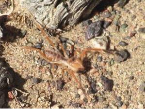
The environmental conditions that these animals prefer are related to the absence of sunlight and dark places, so they tend to spend the day hidden and go out at night to hunt their prey for food..
Solifuges are distinctly carnivorous and are considered one of the most effective predators in the animal kingdom. Their prey are basically represented by other arthropods such as insects, scorpions, spiders and there have even been cases of cannibalism..
Once it identifies a prey, the animal chases and attacks it, especially with its pedipalps, fixing it with the suction cup that they have at their ends. When they capture the prey, with the help of their powerful chelicerae they begin to crush them in order to ingest them..
In these animals, digestion is external, since while they grind their prey with chelicerae, they release digestive juices that contain enzymes. These act on the tissues of the prey, processing and degrading it, turning it into a matter of liquid texture, which is easier to digest completely..
The solífugos reproduce in a sexual way, with a fertilization that can be direct and indirect. In addition, they are oviparous with indirect development.
In the process of reproduction of these animals there may or may not be copulation. When there is copulation, it occurs as follows: the male takes the female and manipulates her until she adopts a position in which the genital pore is easily accessible to the male..
Subsequently, it deposits a drop of his sperm and collects it with his chelicerae, which serve to introduce it into the genital pore for fertilization to occur..
In cases where there is no copulation, the male deposits a spermatophore on the ground, in which the sperm are contained. The female picks it up with her chelicerae and introduces it into the genital pore.
Later, the female lays the eggs (50-100), which have a development period of 12 hours. After this time, the eggs hatch and larvae hatch from them, which undergo a total of six molts until they reach maturity..
The solífugos cover a total of 1100 species approximately, which are distributed in 12 families.
It is the only species found in the Iberian Peninsula. It has scissor-like chelicerae, is small in size (the largest specimen is 3 cm) and has a reddish color. Instead of preferring dark places, it is common to find it in open and clear habitats.
This species is characterized by its very well developed chelicerae, its wider-than-normal opistosome and the large number of sensitive extensions (hairs) that cover its entire body. They are widely distributed throughout southern Africa.
It is a species exclusive to Ethiopia. Their body color is light and their legs are usually very long. They are large in size compared to other species of solifuge, and their chelicerae are widely developed.


Yet No Comments