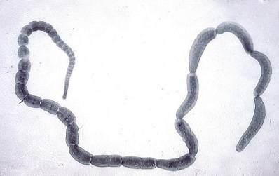
Dipylidium caninum It is an animal that belongs to the class Cestoda of the phylum of the flatworms and that presents the classic morphology of these; a flattened and segmented body.
It has been known for quite some time, having been described for the first time by the famous Swedish naturalist Carlos Linnaeus. However, who deepened his study was the so-called father of parasitology, the German Karl Leuckart.
This parasite is widely distributed throughout the world and to infect its hosts, it requires the flea as an intermediary, which is why most of its definitive hosts tend to be cats and dogs..
Article index
Dipylidium caninum it is an organism classified within the multicellular eukaryotes. This is so because their cells have a cell nucleus, within which are the chromosomes, made up of DNA. They are also composed of different types of cells, specialized in specific organic functions..
This parasite is triblastic, since during its embryonic development the three germ layers are present: ectoderm, mesoderm and endoderm. They are also acellomed, that is, they do not have an internal cavity (coelom).
The lifestyle of Dipylidium caninum it is a parasite, so in order to survive it requires being inside a host, which in most cases is a mammal such as a cat or dog. Man can also be a guest at times.
This parasite is hermaphroditic, presenting female and male reproductive organs. They are oviparous animals, since they reproduce through eggs. They are also pathogenic, since they are the causative agents of dipylidiasis.
Like all animals that belong phylum Platyhelminthes, Dipylidium caninum It has a dorsoventrally flattened shape and three segments: head, neck and body. Its size is regular, generally about 30 cm. However, specimens have been obtained that have measured up to 70 cm in length.
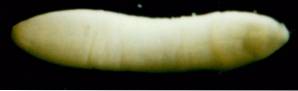
Their color is variable, although they are always light colors such as yellow, cream or white..
Known by the name of scolex, it is more bulky compared to the rest of the body. It is rhomboid in shape and has a rostellum that is apical, conical, and retractable that can have several rows of hooks. These are variables, and there may be from 1 to 6 approximately.
In addition to this it has four suction cups.
The body is made up of several segments called proglottids. Each of these has two pores and has an elongated ovoid shape in the longitudinal direction. They measure between 10 and 12 mm in length. The approximate number of proglottids that an adult worm can have ranges between 50 and 150.
Inside the proglottids there are both male and female reproductive organs. Similarly, there are two types of proglottids: immature and gravid. The immature are those that are closer to the neck and head, they are not yet mature from the sexual point of view.
The proglottids closest to the terminal end of the parasite are sexually mature, which implies that they may be laden with eggs. That is why they are known as gravid proglottids. These are shedding the parasite and are expelled to the outside with the feces or even by themselves.
The taxonomic classification of Dipylidium caninum is the next:
-Domain: Eukarya
-Animalia Kingdom
-Phylum: Platyhelminthes
-Class: Cestoda
-Order: Cyclophyllidea
-Family: Dipylidiidae
-Gender: Dipylidium
-Species: Dipylidium caninum
The life cycle of Dipylidium caninum It is somewhat complex, since it contemplates the intervention of two intermediate hosts, such as the flea and some mammal such as the dog or the cat..
It is important to remember that the Cestoda class worms have proglottids, some of which are gravid, that is, they contain a large number of eggs, protected by an embryonic cover..
These proglottids are released into the environment by two mechanisms. They can be dragged in the stool, in the form of small chains and also come out through the anus spontaneously.
Once exposed to environmental conditions, the proglottids undergo a disintegration process and release the eggs that are contained in them. There in the environment are found the larvae of the intermediate host, the flea.
The flea larvae, which can be those that affect cats or dogs, ingest the eggs. For this process to be successful, it is essential that the flea is in its larval stage, since when it reaches adulthood, its digestive structures do not allow the ingestion of solid particles.
Inside the flea, the parasite undergoes a transformation and becomes the oncosphere, which is the next larval stage. Oncospheres are characterized by having a spherical shape and presenting cilia around them, as well as having hook-like structures, which allow them to penetrate the intestinal wall of their host..
There, it continues its development and reaches the next stage, which is that of cysticercoid. It is worth mentioning that this is the infective stage of this parasite, so if it is ingested by its definitive host (mammal), it can infect it..
Definitive infection occurs when fleas that are infected by cysticercoids are ingested by the animal, primarily a dog. Once inside this host, cysticercoids travel through the digestive tract until they reach the small intestine..
Here, the parasite, with the help of the specialized structures found in its cephalic portion, anchors itself to the intestinal wall and begins to feed on the nutrients that its host ingests..

Thanks to this, the parasite successfully completes its development and reaches sexual maturity, then begins to produce proglottids that contain a large number of eggs inside..
Later, as with the rest of the cestode parasites, the terminal proglottids begin to detach and be expelled through the host's anus to start the cycle again..
Humans can be an incidental part of the cycle when fleas infected with cysticercoids are accidentally ingested. This is more common than is believed, especially among infants, since as the dog is a domestic animal, they tend to handle them and come into contact with the feces of these animals..
Dipylidium caninum it is the parasite responsible for a disease known as dipylidiasis, which is common among domestic animals such as cats and dogs, although it also affects humans.
This parasite has an approximate incubation period of between 3 and 4 weeks. That is the time it takes for the parasite to become an adult and begin to produce eggs..
As already explained, this parasite enters its hosts through the ingestion of fleas that contain within them the larval stage of the parasite called cysticercoid. Dogs and cats can ingest it by licking their fur. While humans can do it when handling their pets.
Person-to-person contagion is totally ruled out.
In general, infection by Dipylidium caninum It can be asymptomatic, so there are no warning signs that warn of the presence of this parasite during its early phase.
However, as the parasite takes hold and anchors in the intestine of its host, it begins to cause certain discomforts that eventually translate into certain symptoms. Because it is an intestinal parasite, the main symptoms affect the digestive tract. These include:
-Epigastric pain
-Occasional diarrhea
-Flatulence
-Constipation
-Abdominal distension
-Vomiting
-Sickness
-Loss of appetite
-Itching at the anal level, generated by the presence of the proglottids in this area.
-Pain in the anal opening.
-Involuntary weight loss, due to the parasite feeding on the nutrients that its host ingests.
There are also other signs and symptoms that derive from the discomfort caused by this parasitosis, such as:
-Insomnia
-Irritability
-Decay
-Tiredness
-Restlessness
As with most intestinal parasites, the definitive diagnosis is made by direct observation of the eggs or proglottids in the feces of the infected person..
When the doctor suspects that a patient is infected with an intestinal parasite, the exam he performs is an analysis of feces, which seeks to identify if there are eggs in them, in order to later make a differential diagnosis.
In the case of Dipylidium caninum, proglottids are seen in the stool. These should undergo a histological analysis to be able to observe the egg packets inside and in this way to be able to confirm the infection by this parasite..
The treatment scheme for infections by Dipylidium caninum it is quite simple, using an anthelmintic drug known as praziquantel.
This medicine has several mechanisms of action that neutralize parasites. First, it acts at the cell membrane level, altering the flow of ions such as calcium. This results in the parasite's musculature being affected, causing problems in contraction and relaxation..
What praziquantel does is generate a muscle spasm in the parasite that causes it to be unable to move and ends up dying.
It is worth noting that three months after taking the treatment, it is important to have a new stool test, to be able to check if the infection was controlled.


Yet No Comments