
The term acinetopsia or akinetopsia comes from the Greek words akinesia (absence of movement) and opsis (to see). It was introduced shortly before 1991 by the British neurobiologist Semir Zeki to name a selective deficit in the ability to perceive movement.
Acinetopsia is also known as motion blindness, and it is a neuropsychological disorder in which a patient cannot perceive movement in his visual field, despite being able to see stationary objects without problem. There are varying degrees of acinetopsia - from viewing motion like a film reel to the inability to discriminate any type of motion. There is currently no effective treatment or cure for acinetopsia..
Contents
Acinetopsia can be separated into two categories based on the severity of the symptom and the amount of blindness affecting the patient.
In these cases the movement is perceived as a film reel or a multiple exposure photograph. This is the most common type of acinetopsia and many patients consider this type of strobe viewing a huge nuisance. This acinetopsia often occurs with afterimages left in each frame of movement..
The pathophysiology of this type of acinetopsia is not known, but it has been hypothesized that it is due to inappropriate activation of physiological movement suppression mechanisms that are normally used to maintain visual stability during eye movements (for example, saccadic suppression).
Gross acinetopsia is an extremely rare disorder. Patients have profound motion blindness and enormous problems carrying out activities of daily living. Rather than viewing the vision as a film reel, these patients have trouble perceiving the movement in its entirety. Most of what is known about this extremely rare condition was learned through the case study of one patient, LM. He described pouring a cup of tea or coffee as difficult "because the liquid appeared to be frozen, like a glacier." She didn't know when to stop pouring, because she couldn't feel the movement of the liquid rising into the cup. LM and other patients have also expressed having trouble following conversations, because lip movements and changing facial expressions were lost. LM stated that she felt unsafe when more than two people walked through the room: "people were suddenly here or there but I hadn't seen them move." Movement is deduced by comparing the change in position of an object or person. LM and others affected have described that crossing the street and driving a car is almost impossible. LM to deal with these problems, he trained his hearing to be able to estimate the distance of moving objects.

The causes of acinetopsia can be: the interruption of the cortical area that is in the central zone of the temporal lobe, it can appear as a secondary effect to certain antidepressant drugs, it can also be caused by a cerebral infarction or by certain cranial surgeries. In some cases, acinetopsia can go away when antidepressant treatment is stopped or through brain surgery..
A change in the structure of the brain (usually injuries) is capable of altering the psychological process of understanding sensory information, in this case visual information. Alteration of visual movement only is possible due to the anatomical separation of visual movement processing from other functions. Like acinetopsia, the perception of color can also be selectively altered as in the case of achromatopsia (also called monochromatism), which is a congenital and non-progressive disease that consists of an abnormality of vision as a result of which only the colors white, black, gray and all their tonalities are perceived.
There is an inability to see movement despite normal spatial acuity. Other intact functions include the perception of visual space and the visual identification of shapes, objects, and faces. In addition to simple perception, acinetopsia also impairs visual-motor tasks, such as reaching for objects and catching objects. When performing tasks, feedback from one's own movement appears to be important.
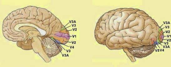
Acinetopsia may be a deficit acquired by lesions on the posterior side of the visual cortex (area V5). These types of lesions most often cause gross acinetopsia. Neurons in the medial temporal cortex respond to mobile stimuli and is therefore the movement processing area of the cerebral cortex. In the aforementioned case of SCI, for example, the brain lesion was bilateral and symmetrical, and at the same time small enough not to affect other visual functions. Some unilateral injuries have also been reported to affect perception of movement. Acinetopsia through lesions is rare, as damage to the occipital lobe generally disrupts more than one visual function.
Acinetopsia can be triggered by high doses of certain antidepressants and vision returns to normal once the dose is reduced.
Regular use of hallucinogenic drugs can also lead to severe perceptual disorder and even motion blindness, although the least severe, acinetopsia fina.
Migraine is a severe headache that usually affects one side or part of it and is often accompanied by nausea and vomiting. It can present very varied symptoms. The most frequent are neurological, gastrointestinal and sensitive..
The aura is a phenomenon mainly of visual origin, although not exclusively, that appears a few hours or minutes before the attack. It is like a warning of the imminent arrival of pain. They are transitory episodes of variable duration, usually lasting between four and 60 minutes. It appears to be due to the small contractions of the vessels before dilation in response. During these contractions, the blood supply to some areas of the brain is temporarily decreased; but it is enough for the characteristic signs of this state to appear. It is this decrease in irrigation that produces the appearance of: visual symptoms, such as dots, flashes, rays, fractional or mosaic images, decreased visual field ... these being the most frequent signs of aura and acinetopsia.
Fine or discrete akinetopsia can be selectively and temporarily induced by transcranial magnetic stimulation (TMS) of the V5 area of the visual cortex in healthy subjects. It is carried out on a surface of 1 cm² of the head, which corresponds in position to area V5.
In addition to memory problems, Alzheimer's patients can suffer from varying degrees of acinetopsia. This could contribute to their marked disorientation. While Pelak and Hoyt have recorded a case of Alzheimer's, not enough research has yet been done on the subject.
Zihl, J., Cramon, D., Mai, N. (1983). Selective alteration of movement vision after bilateral brain damage. Brain, 106, 313-340.
Zeki, S. (1991). Acinetopsiacerebral (visual movement blindness). Brain, 114, 811-824.


Yet No Comments