The acholia It is the lack of coloration of the stool due to the lack of bile pigments in its structure. The English equivalent, acholia, refers more to the absence of bile secretion than to fecal discoloration, but they do state that one of the consequences of acholia is the expulsion of pale or white stools.
Its etymology is very simple: the prefix “a-” means “without” or “lacking in”, and the rest of the word, colia, refers to bile and not color, as may be believed by their similarity in what is written and what is spoken. It would be literally translated as "without bile" or "devoid of bile".
There are several causes of acholia, all of which are related to a lack or decrease in the production and release of bile into the duodenum. The main cause is the obstruction of the bile ducts, basically the common bile duct. Treatment will depend on the cause, and may be surgical or medical.
Article index
It is the blockage or cessation of bile flow, which prevents bile from reaching the small intestine, specifically the duodenum.
In addition to acholia, cholestasis presents with coluria, jaundice, and severe itching. This condition is divided into two large groups, depending on the level of the obstruction or the origin of the problem:
In this type of cholestasis, the damage that causes it occurs directly in the liver or the obstructed bile ducts are still within the liver parenchyma. There are pathologies that cause acute or chronic intrahepatic cholestasis, among which are:
- Viral hepatitis.
- Toxic hepatitis.
- Postoperative benign cholestasis.
- Liver abscesses.
- Biliary atresia.
- Caroli's disease.
- Byler's disease.
- Arteriohepatic dysplasia.
- Alpha-1-antitrypsin deficiency.
- Sclerosing cholangitis.
- Biliary cirrhosis.
- Cholangiocarcinoma.
- Autoimmune hepatitis.
- Sarcoidosis.
- Amyloidosis.
- Heart failure.
- Cholestasis of pregnancy.
- Hodgkin's disease.
- Recurrent benign cholestasis.
In this case there is no direct damage to the liver, but rather an exogenous obstruction of the bile ducts due to different causes, including the following:
- Gallstones (choledocholithiasis).
- Tumors in the head of the pancreas.
- Bile duct cancer.
- Cholangitis.
- Pancreatitis.
- Common bile duct cysts.
- Biliary ascariasis.
Drug-induced hepatotoxicity represents up to 40% of cases of liver failure caused by drugs, and its consequences include compromised bile flow and acholia.
There are many drugs capable of inducing liver damage, so only the most important are mentioned by group:
- Cephalosporins.
- Macrolides.
- Quinolones.
- Penicillins.
- Chlorpromazine.
- Haloperidol.
- Barbiturates.
- Sertraline.
- Diclofenac.
- Ibuprofen.
- Meloxicam.
- Celecoxib.
- Captopril.
- Irbesartan.
- Methyldopa.
- Diuretics.
- Clopydrogrel.
- Warfarin.
- Glimepiride.
- Metformin.
- Steroids.
- Statins.
- Ranitidine.
- Cyclophosphamide.
- Parenteral nutrition.
Bile, commonly known as gall, is produced by the liver and stored in the gallbladder. This substance not only fulfills digestive tasks, helping with the emulsion of fatty acids, but also helps with the transport and elimination of certain waste products.
This last task is important when it comes to the degradation of hemoglobin. The final elements when hemoglobin separates are globin and the "heme" group, which finally transforms into bilirubin and iron after being subjected to a series of biochemical processes in the liver..
Bilirubin is initially found outside the liver in its unconjugated or indirect form. Transported by albumin, it reaches the liver where it binds to glucuronic acid, conjugates and accumulates later in the gallbladder. There it joins with other elements such as cholesterol, lecithin, bile salts and water, to form bile..
Once bile is formed and stored, certain specific stimuli are expected for its release. These stimuli are usually the intake of food and its passage through the digestive tract. At that time, the bile leaves the gallbladder and goes to the duodenum, through the bile ducts and the common bile duct..
Once in the intestine, a certain percentage of the bilirubin that makes up the bile is transformed by the intestinal flora into urobilinogen and stercobilinogen, colorless and water-soluble compounds that follow different pathways. Stercobilinogen is oxidized and becomes stercobilin, which gives the stool a brown or orange hue..
This whole process can be altered when the production of bile is insufficient or when its release is partially or totally limited by an obstruction of the bile ducts..
Acholic stools
If the bile does not reach the duodenum, the bilirubin is not transported to the small intestine and the action of intestinal bacteria on it is not possible..
Due to this, there is no production of stercobilinogen and less of its oxidation product, stercobilin. As there is no element that stains the stool, they are expelled colorless or pale.
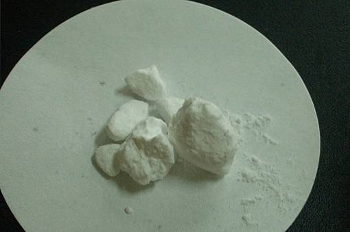
The authors give different shades to acholic stools. Some describe them as pale, clay-colored, putty, clear, chalky, or simply white..
What is significant about all this is that acholic stools will always be related to a disorder in the production or transport of bile, being a very guiding clinical sign for the doctor..
To eliminate acholia, the cause of it must be treated. Among the therapeutic alternatives are medical and surgical.
Choledochal stones are often resolved through lower digestive endoscopies, but those that accumulate in the gallbladder require surgery..
The most common operation is cholecystectomy or removal of the gallbladder. Some tumors can be operated on to restore bile flow, as well as local strictures and cysts.
Infectious causes of cholestasis, acute or chronic, should be treated with antimicrobials. Liver and bile abscesses are often caused by multiple germs, such as bacteria and parasites, so antibiotics and anthelmintics can be helpful. Penicillins, nitazoxanide, albendazole and metronidazole are of choice.
Autoimmune and deposit pathologies are usually treated with steroids and immunomodulators. Many cancer diseases that cause cholestasis and acholia should be treated initially with chemotherapy, and then possible surgical alternatives should be considered.

Yet No Comments