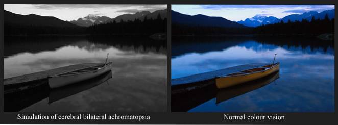
The achromatopsia It is a vision defect in which the person who suffers from it is not able to distinguish colors. Condition also known as monochrome vision, it is characterized by perceiving only the colors white, gray and black, as well as their tonalities.
The ability to not distinguish colors in patients with achromatopsia can be total or partial. In addition, they present other problems such as decreased visual acuity, involuntary movements of the eyes or nystagmus, sensitivity to light or photophobia and impossibility for fixation of vision to a point.
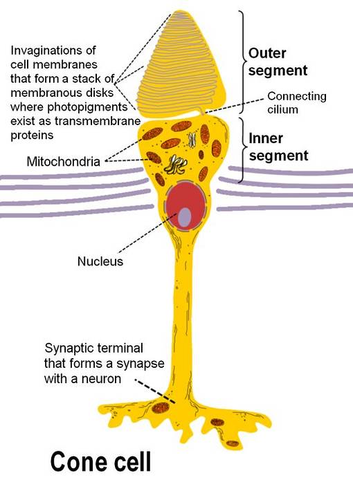
This inability for color distinction can be genetic or acquired. When it occurs from birth due to genetic abnormalities, the problem is in the color perception cells located in the eyes called cones.
On the contrary, in the case of an acquired condition, the problem is found centrally, in the signal transmission pathways from the eyes to the brain, frequently as a consequence of trauma or ischemic vascular disease. These patients do not have eye dysfunctions.
The treatment of this condition is based on measures to improve the quality of life of the patient, since there is no cure.
Article index
The causes of achromatopsia can be genetic or acquired. If they are genetic, they appear from birth, being a rare condition, since it is a genetic mutation that only occurs in 1/30,000 individuals. In the case of being acquired, the underlying disease must damage the specific part of the cerebral cortex that interprets colors.
Patients with genetic achromatopsia have a dystrophy in the cells of the eyes that are responsible for perceiving colors and sending signals in the form of electrical impulses to the brain where they are interpreted. These cells are called cones and are located in the retina.
The problem in the cones is mediated by specific genes that act at this level during the formation in the fetus.
There are 3 types of cones: those that are sensitive to the color red, those that are sensitive to the color blue and those that are sensitive to the color green. The type of dysfunction that the patient has will depend on the group of cones that is atrophied.
The most common is that the person is unable to distinguish all the colors, therefore they will have a vision in black, black and gray scale. This type of achromatopsia is called complete achromatopsia or typical.


There is also a partial or incomplete type, atypical, in which the patient cannot distinguish a specific color.
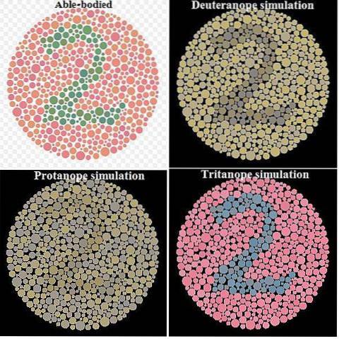
The partial type takes on specific names to refer to each condition. Thus, the dystrophy of green perception cones is called: deuteranotopia; the one with red perception cones is the protanotopia and the blue perception cones tritanotopia.
Acquired achromatopsia is secondary to an external cause that causes damage to the cerebral cortex, specifically in the part specialized in interpreting colors.
It can occur due to severe head trauma, but is usually the result of ischemic cardiovascular disease that causes decreased or absent vascularization in that brain area..
These patients have no eye problems and their vision is normal until the time of the accident that caused the brain damage..
In this type of achromatopsia the symptoms are different from those found in a patient with genetic disease. It is often accompanied by other perceptual disorders such as the inability to recognize familiar faces or prosopagnosia.
Patients with achromatopsia present from an early age involuntary movements of the eyes in a horizontal plane, called nystagmus; also decrease in visual acuity, which is the sharpness with which images are observed in adequate lighting conditions.
They are also very sensitive to light, presenting the disorder called photophobia and may have blurred vision under very bright lighting or hemeralopia.
The inability to identify the colors can be partial or total, but the most common is that it is complete and the person perceives all the colors in the gray scale.
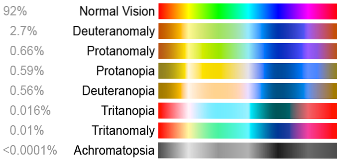
In partial achromatopsia, the patient presents all the symptoms of his total counterpart, but with less intensity.
The diagnostic approach can be made by the specialist in a child with nystagmus, or eye movement, when other causes of this symptom are ruled out..
Later, a complete ophthalmological evaluation should be performed in which visual acuity and color perception are examined..
To assess a person's ability to distinguish colors, a test is used in which a series of cards with specific designs for this purpose is observed. They are called Ishihara letters.
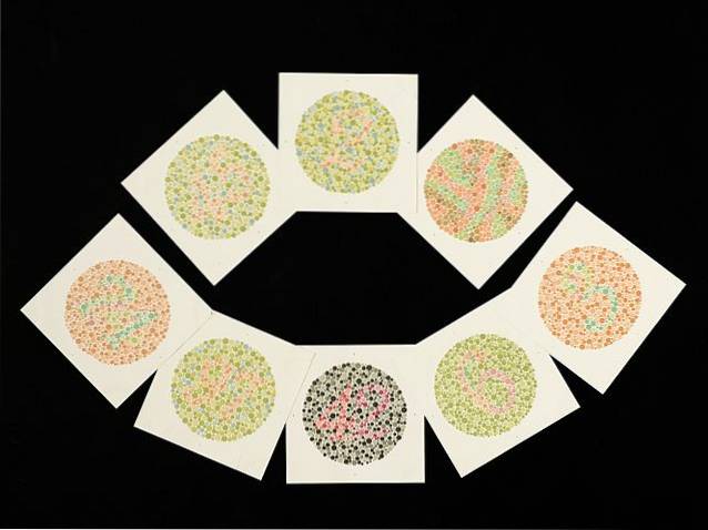
The cards were designed by the Japanese ophthalmologist Shinobu Ishihara in 1917. The drawing consists of a circular image that in turn contains small colored circles inside, which draw a number on the red and blue scales..
The card game consists of 38 cards, but the clutter is usually quickly detected when starting the test.
The definitive diagnosis of achromatopsia is made from a genetic test that reveals the mutation.
Currently there are no treatments to cure achromatopsia, although there are studies in the experimental phase in which intraocular injections of specific factors that help to regenerate the activity of the cones are carried out..
Patients with achromatopsia present bothersome symptoms such as photophobia and hemeralopia, which is why the use of contact lenses with special filters is indicated to improve their vision during the day..
Visual acuity problems improve with the use of lenses with specific formulas for each case..
Children with achromatopsia should go to a specialized consultation every 6 months and adults between 2 and 3 years.
Despite the proper application of these treatments, patients with difficulty distinguishing colors have problems performing common activities such as driving and attending a class at school..
Genetic counseling with a specialist is recommended for people who have the disease, or whose parents have it, at the time of family planning. This will explain the risks and probabilities of having a child with the condition..


Yet No Comments