The Silvio aqueduct, Also known as the cerebral aqueduct or the midbrain aqueduct, it is a communicating region of the brain. This structure is characterized by connecting the third cerebral ventricle with the fourth cerebral ventricle and its main function is to allow the circulation of cerebrospinal fluid.
Silvio's aqueduct is located posterior to the bridge and limits caudally with the medulla oblongata and the cerebellum. It is not a functional brain structure, it simply acts as a communication aqueduct between different brain regions. However, alterations in its functioning have been related to important pathologies.

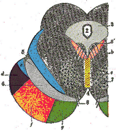
Silvio's aqueduct is located between the midbrain and the metancephalon. In its dorsal region is the brainstem bridge and in the ventral region the cerebellum.
Likewise, it is part of the ventricular system of the brain, develops from the central canal of the neural tube and originates in the region of the neuronal tube that is present in the developing midbrain..
Article index
The aqueduct of Silvio refers to what is known today in medical terms as the aqueduct of the midbrain or cerebral aqueduct.
In the field of medicine, Silvio's aqueduct terminology has fallen into disuse, however, since it is the original name, many manuals and review articles still call it that way..
As its name suggests, the Silvio aqueduct is a cerebral aqueduct. That is, a structure that connects two different regions of the brain. Specifically, it connects the third and fourth ventricles of the brain.
However, the aqueduct of Silvio plays a more important role than the connection between ventricles, since it is the region of the brain that allows the circulation of cerebrospinal fluid.
Cerebrospinal fluid is a colorless fluid that bathes the brain and spinal cord. This fluid performs important actions in the brain such as cushioning trauma or providing hydropneumatic support to the brain..
The ventricular system consists of a series of cavities in the brain that develop within the central nervous system. These regions are mainly responsible for producing and allowing the circulation of cerebrospinal fluid..
The regions that are part of the ventricular system are the lateral ventricles, the third ventricle, the aqueduct of Silvio and the fourth ventricle..
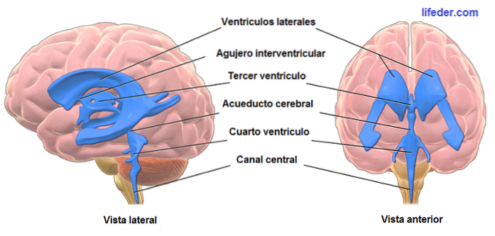
The lateral ventricles are found in each cerebral hemisphere, they are shaped like the letter "C" and each of them contains a posterior horn, an anterior horn and a lower horn..
The lateral ventricles communicate with the third ventricle through the interventricular foramen or foramen of Monroe.
The third ventricle is a cleft-shaped region of the brain. It is located in the midline between the right thalamus and the left thalamus, and the right hypothalamus and the left hypothalamus.
The third ventricle connects with the lateral ventricles, as well as with the fourth ventricle thanks to the aqueduct of Silvio.
The aqueduct of Silvio or cerebral aqueduct is a narrow conduit that measures approximately 18 millimeters in length. This is located between the third and fourth ventricle, allowing the connection between both and transporting the cerebrospinal fluid from and to these structures..
Finally, the fourth cerebral ventricle is a cavity that is located between the brainstem and the cerebellum. The roof of the fourth ventricle borders the cerebellum, while the foot is formed by the posterior aspect of the bridge and the medulla oblongata.
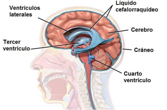
Cerebrospinal fluid (CSF), also known as cerebrospinal fluid (CSF), is a colorless fluid that bathes the brain and spinal cord. It circulates through the subarchnoid space, the cerebral ventricles and the ependymal canal. This liquid is a basic substance for the functioning of the brain.
Specifically, the CSF keeps brain tissue floating, acting as a cushion, serves as a vehicle to transport nutrients to the brain and eliminate waste, and flows between the skull and spine to compensate for changes in intracranial blood volume.
CSF is formed in the choroid plexuses of the four cerebral ventricles. Its circulation begins in the lateral ventricles and continues to the third ventricle through the foramina of Monroe.
Once the CSF reaches the third ventricle, the aqueduct of Silvio comes into play, since it is this brain structure that allows the continuation of CSF transport to the fourth ventricle..
Once the CSF reaches the fourth cerebral ventricle, it is conducted through a set of orifices to the cisterna magna, a large reservoir of fluid located behind the medulla oblongata.
The disease that is related to the functioning of the aqueduct of Silvio is hydrocephalus, a pathology that originates due to an abnormal increase in the volume of cerebrospinal fluid within the brain.
This pathology is usually accompanied by intracranial hypertension and may be due to different causes such as: abnormal increase in CSF production, blockage in CSF circulation or decreased CSF absorption..
At present, different types of hydrocephalus have been described and one of them, communicating hydrocephalus, originates due to an obstruction of the CSF in the aqueduct of Silvio.

Regarding the etiology of hydrocephalus, it has now been established that it can be congenital or acquired. When acquired, it can be due to different factors: infections, hemorrhages or vascular malformations that compromise the aqueduct of Silvio.
In this sense, some cases of hydrocephalus can be operated on by removing the obstruction by dilating the aqueduct of Silvio with stenosis..


Yet No Comments