The blood agar It is a solid enriched, differential but non-selective culture medium. It is used for the recovery and growth of a great variety of microorganisms from clinical samples or for subcultures..
Classic blood agar should be included for the seeding of most clinical samples received in the laboratory; except for stool samples where it is not useful, unless prepared with certain modifications.
This culture medium basically consists of an enriched base agar and 5% blood. The agar base can vary according to the needs, but it will mainly be composed of peptones, amino acids, vitamins, meat extract, sodium chloride, agar, among others..
As for blood, it is normally required to have contact with a livestock facility to obtain blood from animals, such as sheep, rabbit or horse. However, this is not always possible and human blood is sometimes used..
Blood agar medium can be prepared in the laboratory or can be purchased ready-made from dedicated companies. The preparation of this medium is one of the most delicate, any carelessness in its preparation will result in a contaminated batch.
That is why all possible precautions must be taken and in the end a quality control must be carried out incubating at 37 ° C 1 plate for every 100 that are prepared.
Article index
Blood agar is an enriched medium because it contains 5-10% blood on an agar base as its main additive. Both compounds contain many nutrients and this property allows most cultivable bacteria to grow in it..
That growth occurs without restriction; for this reason it is non-selective. However, if compounds are added to this medium that prevent the growth of some microorganisms and favor that of others, it becomes selective. This is the case if certain types of antibiotics or antifungals are added.
Likewise, blood agar is a differential medium, since it allows us to distinguish 3 types of bacteria: beta-hemolytic, alpha-hemolytic and gamma-hemolytic..
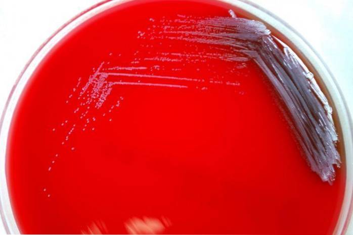
Beta-hemolytics are those that have the ability to completely lyse or break red blood cells, forming a clear halo around the colonies, therefore they produce ß or ß-hemolysis and the microorganisms are called ß-hemolytics..
Examples of ß-hemolytic bacteria are Streptococcus pyogenes Y Streptococcus agalactiae.
Alpha-hemolytics are those that carry out partial hemolysis, where hemoglobin is oxidized to methemoglobin, generating a greenish coloration around the colonies. This phenomenon is known as α-hemolysis or α-hemolysis and bacteria are classified as α-hemolytic..
Examples of α-hemolytic bacteria are Streptococcus pneumoniae Y Streptococcus of the group viridans.
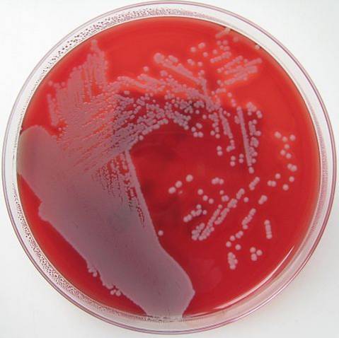
Finally, there are the so-called gamma-hemolytic or non-hemolytic bacteria. These grow on the agar without generating changes on it, an effect known as γ-hemolysis, and the microorganisms are γ-hemolytic.
Example of γ-hemolytic bacteria: some strains of group D Streptococcus (Streptococcus bovis and Enterococcus faecalis).
The blood agar culture medium is one of the most commonly used in the microbiology laboratory..
Among the microorganisms capable of growing in the blood agar medium are: strict aerobic, facultative, microaerophilic, anaerobic, Gram positive or Gram negative bacteria, fast growing or slow growing bacteria.
Some nutritionally demanding or fastidious bacteria also grow, as well as fungi and yeasts. Likewise, it is useful to perform subcultures or reactivate strains that are metabolically very weak..
However, the choice of blood type and base agar will vary depending on the probable microorganism suspected of recovering and the use that will be given to the plate (culture or antibiogram)..
Blood can be lamb, rabbit, horse or human.
The most recommended is lamb's blood, with some exceptions. For example, to isolate Haemophilus species, where the recommended blood is horse or rabbit blood, since lamb blood has enzymes that inhibit factor V.
The least recommended is human, however it is the most used, perhaps because it is the easiest to obtain.
The blood must be defibrinated, obtained without any type of additive and from healthy animals. For the use of human blood, several factors must be taken into account:
If the blood comes from individuals who have had bacterial infections, they will have specific antibodies. Under these conditions, the growth of some bacteria is likely to be inhibited..
If it is obtained from the blood bank, it contains citrate and certain bacteria may not grow in its presence. On the other hand, if the blood comes from patients taking antibiotics, the growth of susceptible bacteria can be inhibited..
And if the blood is from a diabetic person, excess glucose interferes with the proper development of hemolysis patterns.
The base agar used for the preparation of blood agar can be very broad. Among them are: nutrient agar, brain heart infusion agar, trypticase soy agar, Müeller Hinton agar, Thayer Martin agar, Columbia agar, Brucella agar, Campylobacter agar, etc..
This base is the least used, since it will mainly grow non-demanding bacteria, such as enteric bacilli., Pseudomonas sp, S. aureus, Bacillus sp, among others. It is not recommended to isolate Streptococcus.
It is one of the most used as a base for blood agar, because it has the necessary nutrients for the growth of most bacteria, including Streptococcus sp and other fastidious bacteria. Although not suitable for observing patterns of hemolysis.
Lamb's blood is generally used with this base.
Blood agar variants can also be prepared, where other compounds are added to isolate certain microorganisms. For example, brain heart infusion agar supplemented with rabbit blood, cystine and glucose, serves to isolate Francisella tularensis.
Whereas, with cystine tellurite it is useful for the isolation of Corynebacterium diphteriae. Human or lamb blood can be used.
With the first beta-hemolysis will be seen as a narrow halo, while with the second the halo will be much wider.
Likewise, this base together with bacitracin, corn starch, horse blood and other enrichment supplements (IsoVitaleX), is used for the isolation of the genus Haemophilus sp from respiratory samples.
Also, if the combination of antibiotics chloramphenicol - gentamicin or penicillin - streptomycin with horse blood is added, it is ideal for the isolation of demanding pathogenic fungi, even with a higher yield than Sabouraud glucose agar. It is especially useful in isolating Histoplasma capsulatum.
This base is the most recommended for a better observation of the hemolysis pattern and for performing diagnostic tests such as optoquine taxa and bacitracin. It is the classic blood agar that is used routinely.
With this base you can also prepare the special blood agar for Corynebacterium diphteriae, with cystine tellurite Y lamb's blood.
Likewise, the combination of this agar with lamb's blood, plus kanamycin-vancomycin is ideal for the growth of anaerobes, especially Bacteroides sp.
This base supplemented with blood is used to perform the antibiogram of demanding microorganisms, such as Streptococcus sp.
It is also useful for the isolation of bacteria such as Legionella pneumophila.
This medium is ideal as a base for blood agar when Neisseria genus is suspected, especially Neisseria meningitidis, as N. gonorrhoeae does not grow on blood agar.
It is also used to perform susceptibility tests to Neisseria meningitidis.
This base is excellent for seeding gastric biopsy specimens for Helicobacter pylori.
The medium is prepared by adding 7% lamb blood defibrinated with antibiotics (vancomycin, trimethoprim, amphotericin B and cefsulodin) to restrict the growth of other types of bacteria that may be present..
This same base supplemented with human or lamb blood, nalidixic acid and colistin is useful to isolate Gardnerella vaginalis. Also ideal for assessing antimicrobial susceptibility to antibiotics of the same microorganism.
In addition, it is used for the preparation of blood agar for the cultivation of anaerobes, adding aminoglycosides and vancomycin..
This base allows you to properly observe the hemolysis patterns.
This medium used as a base for blood agar together with the addition of vitamin K is ideal for the cultivation of anaerobic bacteria. In this case, the use of lamb's blood is recommended..
Campylobacter agar supplemented with 5% sheep blood and 5 antibiotics (cephalothin, amphotericin B, trimethoprim, polymyxin B and vancomycin), is the medium used to isolate Campylobacter jejuni in stool samples.
Each commercial house brings the instructions to prepare a liter of culture medium on the back of the container. The corresponding calculations can be made to prepare the desired amount, depending on the selected base agar.
The base agar is dehydrated (powder), therefore it must be dissolved in distilled water adjusted to pH 7.3.
The amount indicated by the chosen base agar is weighed and dissolved in the corresponding amount of water in a flask, then heated over moderate heat and mixed with rotary movements until all the powder is dissolved..
Once dissolved, sterilize in an autoclave at 121 ° C for 20 minutes.
When leaving the autoclave, the flask is allowed to cool until the temperature oscillates between 40 to 50 ° C; It is a temperature that the human skin supports and at the same time the agar has not yet solidified.
To do this, the flask is touched with the hand and if the heat is tolerable, it is the ideal temperature to add the corresponding amount of defibrinated blood (50 ml for each liter of agar). Mix gently to homogenize.
The passage of blood aggregation is crucial, because if it is done when the medium is very hot the red blood cells will break down and the medium will not serve to observe hemolysis.
Adding too cold will cause lumps to form and the surface of the media will not be smooth to allow proper scoring..
Serve in sterile Petri dishes immediately after homogenizing the blood. Approximately 20 ml is poured into each Petri dish. This procedure is performed in a laminar flow hood or near the burner.
When serving the blood agar in the Petri dishes, no air bubbles should remain on the surface of the plate. If this happens, the flame of the Bunsen burner is passed quickly over the plate to eliminate them..
Plates are allowed to solidify and stored in a refrigerator (2-8 ° C) inverted until use. Before using the blood agar plates, they must be tempered (allowed to reach room temperature) to be able to be sown..
The prepared plates last approximately 1 week.

Yet No Comments