The anaphase it is a phase of the division of the nucleus where duplicated chromosomes separate, and chromatids move to opposite poles of the cell. Occurs in both mitosis and meiosis.
Although the processes of mitosis and meiosis are similar in some of their stages, there are considerable differences in these events. The fundamental difference is that in mitosis there is one anaphase and in meiosis two.
Article index
Before describing the anaphase process, it is necessary to know the basic terminology biologists use to describe chromosomes..
Chromosomes are units of DNA (deoxyribonucleic acid) compacted in a highly efficient way. They have the information necessary for an organism to function and develop. Information is organized into elements called genes.
In humans, for example, there are 46 chromosomes in somatic cells. This number varies depending on the species studied. Since we are diploid organisms, we have one pair of each chromosome, and these are known as a homologous pair..
Regarding the structure of a chromosome, we can distinguish the chromatids. These are each of the longitudinal elements of the same, when it is already duplicated. Each chromosome is made up of two chromatids sisters and the region where they join is called the centromere.
The centromere is a key region, since it is responsible for attaching to the achromatic spindle in the process of cell division. In the centromere there is a structure of a protein nature called the kinetochore. The kinetochore is responsible for anchoring the mitotic spindle.
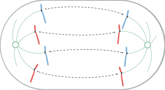
Mitosis is divided into four stages, and anaphase corresponds to the third of these. Includes the separation of sister chromatids, through their simultaneous release from centromeres.
For this to happen, the process is mediated by an enzyme called topoisomerase. The latter is located in the kinetochore region, releases entangled chromatin fibers and facilitates the separation of sister chromatids. Chromosomes move from the centromere at a rate of 1 um per minute.
The central event of anaphase is the separation of the chromatids. This phenomenon occurs thanks to two processes, independent of each other, but coinciding.
One of these is the shortening of the microtubules of the kinetochore, thus the chromatids move further and further away from the equatorial plate towards the poles. In addition, the cell poles are moved away by the elongation of the polar microtubules.
In terms of duration, it is the shortest stage of all mitosis, and it only lasts a few minutes.
At the end of anaphase, each end of the cell has an equivalent and complete set of chromosomes. One of the possible drawbacks in this division phase is the wrong distribution of the two chromatids of a chromosome between the new cells. This condition is called aneuploidy.
To avoid aneuplody, the kinetochore has mechanisms that help to avoid this condition.
Cell division by meiosis is characterized by having two processes or phases of division of the nucleus. For this reason, there is anaphase I and II.
In the first, the centromeres separate and move towards the poles, dragging the two chromatids. The second anaphase is very similar to that found in mitosis.
There are many similarities between the process of division by meiosis and mitosis. For example, in both events the chromosomes contract and become visible under the light of a microscope. However, they differ in several respects.
In mitosis, only one cell division takes place. As is known, the result of mitosis is two daughter cells, genetically the same.
In contrast, meiosis involves two cell divisions, where the product is four daughter cells, different from each other and different from the cell that gave rise to them..
In diploid cells (like ours, with two sets of chromosomes), homologous chromosomes are present before both processes. However, homolog mating only occurs in meiosis.
A crucial difference involved in anaphase is that in meiosis the number of chromosomes is halved in anaphase I.
In this phase of cell division, the separation of homologous chromosome pairs occurs. Note that in mitosis there is no reduction in the genetic load of daughter cells.
One of the most notable characteristics of meiosis is the increase in genetic variation in daughter cells..
These processes are the crossing over and the random distribution of chromosomes from the mother and father. There is no equivalent process in mitotic divisions.
Crossover occurs in prophase I of meiosis, while the random distribution of chromosomes occurs in anaphase I.
Another crucial difference between the two processes is the behavior of chromosomes during anaphase and metaphase..
In metaphase I of meiosis, the alignment of homologous chromosome pairs takes place in the equatorial plane. In contrast, in mitosis those that line up in the aforementioned plane are the individual chromosomes, which corresponds to metaphase II in meiosis..
Then, in anaphase I of meiotic division, the paired chromosomes separate and each of these biological entities migrate towards the poles of the cell. Each of the chromosomes has two chromatids joined by the centromere.
In anaphase of mitosis, and also in anaphase II of meiosis, the sister chromatids separate and each chromosome that migrates towards the poles is made up of only one chromatid..


Yet No Comments