The venous angioma, Technically known as a developmental venous anomaly, it is a group of vascular malformations, considered as a developmental alteration characterized by persisting into adulthood.
This condition usually originates due to alterations in venous drainage during the embryonic stage and stands out as an asymptomatic and benign pathology. Occasionally, venous angioma can cause seizures and, in rare cases, can cause bleeding due to the associated cavernous malformation.
Normally, people with venous angioma do not require treatment and can lead healthy and satisfying lives. However, in some cases, this condition can lead to brain bleeding and relatively intense symptoms..
In recent years, the detection of cases of venous angioma has increased notably due to the diagnostic possibilities presented by new neuroimaging techniques.
Article index
The appearance of venous angioma as a vascular malformation is established in 1951, when Russel and Rubinstein classified these malformations into four main groups.
These groups consisted of telangiectasias, arteriovenous malformations, venous angiomas, and cavernous angiomas..
Years later, in 1963, Courville first described a series of small vascular malformations that consisted solely of venous structures. The main findings about this malformation were:
Later, in 1968 Constants produced the first radiological description of two developmental venous anomalies. Although many authors attribute the first specification of the malformation to Wolf, describing an unusual case of multiple venous angiomas in a subject who died due to intracranial hemorrhage caused by one of these angiomas.
Venous angiomas constitute one of the four cerebral vascular malformations that have been described today. Likewise, the scientific literature shows that it is also the most prevalent of all.
Although it is considered a developmental venous malformation, venous angioma is not exactly an alteration in brain development. In fact, this condition constitutes the persistence in adulthood of an embryonic venous system, so that more than a malformation it should be considered as a variant of normality.
Specifically, although its origin is not well established, several authors point out that it is due to an alteration in the embryonic period that would lead to an occlusion or disorder of the venous drainage system of the brain regions.
In this sense, venous angioma is characterized by presenting a structure composed of small medullary veins that are located deep in the white matter of the brain. These small medullary veins acquire a radial arrangement and converge towards a dilated venous trunk that empties into a normal venous sinus.
The histological architecture of the veins of people with venous angioma is usually similar to that of normal veins and they are surrounded by glial tissue that, in most cases, does not present alterations.
One of the most striking properties of venous angioma lies in the discrepancy between the frequency of this type of brain lesions found in radiological studies and the relatively small number of people with venous angioma.
This fact is mainly due to the fact that the condition is, in most cases, totally asymptomatic.
Thus, most cases of venous angioma are detected when the person undergoes radiological examinations motivated by other conditions or intracranial pathologies, which is why the absence of a diagnosis of this venous anomaly is usually common..
However, it must be taken into account that not all cases of venous angioma are asymptomatic and benign. Occasionally, this abnormality can lead to seizures, headaches, progressive neurological deficits, and bleeding..
The developmental venous anomaly is composed of the convergence of multiple venules with radial arrangement and normal parenchyma between them, which converge in a common trunk collect.
This fact makes the venules referring to venous angioma take on the appearance of a jellyfish and is given the name of Caput medusae..
The venous anomaly can be found in any region of the brain, however, it is usually located in the frontal lobes of the cerebral cortex and in the posterior fossa. Likewise, two thirds of the total venous angiomas found to date are located in the cerebellum.
Venous angiomas are usually characterized by being solitary and unilateral, although some data indicate the existence of bilateral or multiple venous angiomas, especially in the posterior fossa.
Likewise, it must be taken into account that the alteration of the typical drainage of venous angiomas may be different.
For example, in supratentorial angiomas, venous drainage may be superficial. In other words, it can be carried out into cortical veins or dural sinuses. Likewise, in these structures the drainage can also be deep.
Similar drainage pathways are often seen in the posterior fossa of the brain. These pathways include transparenchymal drainage to superficial cerebral veins and dural sinuses, as well as deep drainage to the fourth cerebral ventricle..
The origin of venous angiomas constitutes one of the main challenges for the scientific community today, as it is not completely clear.
Certain authors point out that this anomaly may be motivated by a thrombosis of the drainage vein located in a specific region of the brain that, secondarily, would generate compensatory mechanisms with the opening of embryonic venules that lead to a central trunk.
On the other hand, Saito and Kobayashi suggested in their work the existence of a uterine accident during the formation and development of the medullary and tributary veins, either due to thrombosis or by another mechanism that motivates the formation of a collateral drainage system..
Finally, Padget referred to the possibility that the venous angioma was due to an alteration during pregnancy, a fact that would lead to the formation of compensatory drainage systems.
At present, the three hypotheses have been accepted and the line of research focuses on contrasting or rejecting any of the three. However, none of them has sufficient scientific evidence to be able to establish the etiology of venous angiomas..
In most cases (slightly more than half), venous angiomas are asymptomatic. That is, they do not produce any type of sensation, manifestation or physical and / or neurological complication in the person. However, in some cases this malformation can lead to both specific symptoms and secondary complications..
With regard to symptomatic cases, the most common is that venous angioma presents with headaches and seizures. However, these manifestations may not always be attributable to the radiological findings of venous angioma, since they may have other causes..
On the other hand, people with infratemporal lesions due to venous angioma may experience ataxia and gait disturbances. In this case, the developmental venous abnormality would be considered more of a cause of the brain injury than the pathology that causes the movement symptoms itself..
Another complication that this malformation can lead to is drainage vein thrombosis. This condition can cause non-hemorrhagic and / or hemorrhagic venous infarction. However, it is a very rare complication.
In these rare cases, it has been observed that there is a progressive recanalization of the malformation, and these may bleed spontaneously and causing an increase in intra-arterial pressure..
Despite these complications reported in the venous angioma literature, globally, the risk of bleeding in this type of condition is very low. Specifically, prevalence studies show that these conditions would have an incidence of around 0.22% annually.
On the other hand, several studies show the relationship between venous angioma and cavernous malformation. Data show that at least 30% of cases of developmental venous abnormality could be caused by these factors.
Since most cases of venous angioma are asymptomatic, this developmental abnormality is usually diagnosed in two main ways.
The first (and most prevalent) is usually carried out when the person undergoes radiological studies due to another type of condition and, incidentally, the typical properties of venous angioma are discovered.
The second, on the other hand, is performed during the autopsy, when the pertinent tests detect the presence of the developmental venous anomaly.
Finally, in some cases, venous angioma can be detected when the person presents the typical symptoms of the malformation and it is decided to perform a complete examination to determine the underlying pathology..
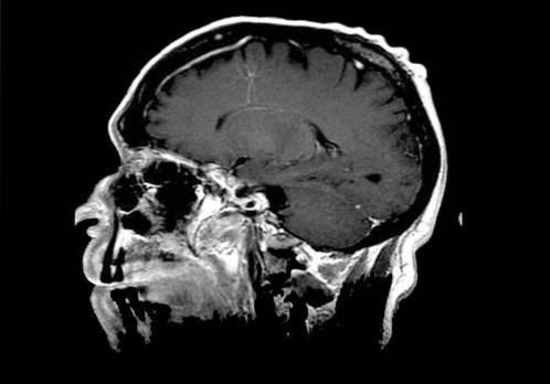
In any of the three cases, computed tomography (CT) is a vital tool for the diagnosis of venous angioma. In fact, without the data on brain anatomy collected by this device, it is impossible to detect the anomaly, so the clinical evaluation alone is insufficient for its diagnosis..
However, conventional computed tomography does not always produce the necessary images to be able to detect anomalies related to venous angioma, which is why the use of high-definition computed tomography is often necessary.
These tools allow the preparation of thin slices and contrast enhancements at the brain level, as well as the reconstruction of computed tomography angiography..
Beyond computed tomography, other devices that can be used for the diagnosis of venous angioma are magnetic resonance imaging (MRI), magnetic resonance angiography (MRA) and conventional angiogram..
Venous angioma is a benign condition in most cases, but in others, it can have negative consequences for the person. In this sense, the main complication in which this developmental venous anomaly can derive is intracranial hemorrhage.
This hemorrhage is usually caused by the obstruction or narrowing of the drainage channel of the lesion, a fact that causes a temporary increase in the pressure of the veins that drain the blood.
Likewise, the most damaging and dangerous element of venous angioma is the role it can play in the generation of other types of vascular malformation with clinical symptoms.
Specifically, developmental venous abnormality has been associated with cerebral cavernous malformation, another type of vascular malformation that often causes seizures, hemorrhages, or focal neurological symptoms..
Likewise, venous angioma has also been related to arteriovenous malformation, a venous malformation that occurs due to an abnormal connection between the arteries and veins of the brain.
This condition usually presents a wide symptomatology, including manifestations such as: confusion, ringing in the ear, headache, trouble walking, seizures, vision problems, dizziness, muscle weakness and body numbness..
The generally passive nature of venous angioma motivates, in most cases, conservative treatment.
In fact, most cases of this vascular anomaly (when it is asymptomatic) do not require any type of treatment, so after the diagnosis of the condition, one must wait for the onset of symptoms before intervening..
In cases where intervention is necessary, evacuation of the intraparenchymal hematoma is recommended, leaving the venous malformation intact. It must be taken into account that surgical intervention for venous angiomas presents high risks of heart attack.
Finally, radiotherapy is not considered indicated to treat this anomaly, since it can induce thrombosis of the malformation and generate serious alterations in the venous drainage of the affected brain region..
Thus, although it is a benign condition in many cases, venous angioma does not currently have effective and safe treatments, so surgical interventions should be avoided whenever possible.


Yet No Comments