The Ribosomal RNAor ribosomal, in cell biology, is the most important structural component of ribosomes. For this reason, they have an indispensable role in the synthesis of proteins and are the most abundant in relation to the other main types of RNA: messenger and transfer..
Protein synthesis is a crucial event in all living organisms. Previously, it was believed that ribosomal RNA was not actively involved in this phenomenon, and that it only played a structural role. Today there is evidence that RNA has catalytic functions and is the true catalyst of protein synthesis.
In eukaryotes, the genes that give rise to this type of RNA are organized in a region of the nucleus called the nucleolus. RNA types are usually classified depending on their behavior in sedimentation, that is why they are accompanied by the letter S for "Svedberg units".
Article index
One of the most striking differences between eukaryotic and prokaryotic lineages is the composition of the ribosomal RNA that constitutes their ribosomes. Prokaryotes have smaller ribosomes, whereas ribosomes in eukaryotes are larger.
Ribosomes are divided into large and small subunits. The small contains a single ribosomal RNA molecule, while the large contains one larger molecule and two smaller ones, in the case of eukaryotes..
The smallest ribosomal RNA in bacteria can be 1,500 to 3,000 nucleotides. In humans, ribosomal RNA reaches greater lengths, between 1800 and 5000 nucleotides.
Ribosomes are the physical entities where protein synthesis occurs. They are composed of 60% ribosomal RNA, approximately. The rest are proteins.
Historically, ribosomal RNA is identified by the sedimentation coefficient of suspended particles centrifuged under standard conditions, which is denoted by the letter S for "Svedberg units.".
One of the interesting properties of this unit is that it is not additive, that is, 10S plus 10S are not 20S. For this reason there is some confusion related to the final size of the ribosomes..
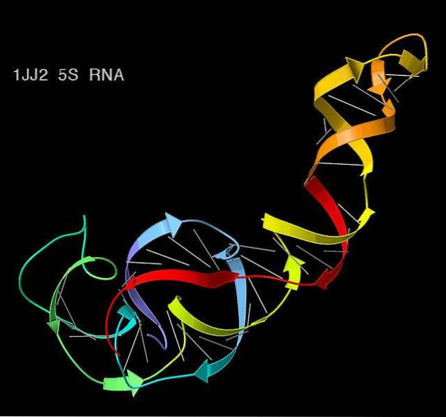
In bacteria, archaea, mitochondria, and chloroplasts, the small unit of the ribosome contains the 16S ribosomal RNA. While the large subunit contains two species of ribosomal RNA: 5S and 23S.
In eukaryotes, on the other hand, 18S ribosomal RNA is found in the small subunit and the large subunit, 60S, contains three types of ribosomal RNA: 5S, 5.8S, and 28S. In this lineage, ribosomes tend to be larger, more complex, and more abundant than in prokaryotes..
Ribosomal RNA is the central component of ribosomes, so its synthesis is an indispensable event in the cell. Synthesis takes place in the nucleolus, a region within the nucleus that is not delimited by a biological membrane.
The machinery is responsible for assembling the ribosome units in the presence of certain proteins.
The ribosomal RNA genes are organized in different ways depending on the lineage. Remember that a gene is a segment of DNA that codes for a phenotype.
In the case of bacteria, the genes for the 16S, 23S, and 5S ribosomal RNAs are organized and transcribed together in an operon. This “genes together” organization is very common in prokaryotic genes..
In contrast, eukaryotes, more complex organisms with a membrane-delimited nucleus, are organized in tandem. In us humans, the genes that code for ribosomal RNA are organized into five “clusters” located on chromosomes 13, 14, 15, 21 and 22. These regions are called NOR.
In the cell, RNA polymerase is an enzyme in charge of adding nucleotides to RNA strands. They form a molecule of these from a DNA molecule. This process of formation of an RNA following as a template a DNA is known as transcription. There are various types of RNA polymerases.
Generally, the transcription of ribosomal RNAs is carried out by RNA polymerase I, with the exception of 5S ribosomal RNA, whose transcription is carried out by RNA polymerase III. The 5S also has the peculiarity that it is transcribed outside the nucleolus.
The promoters of RNA synthesis consist of two elements rich in GC sequences and a central region, here the transcription begins.
In humans, the transcriptional factors necessary for the process bind to the central region and give rise to the pre-initiation complex, which consists of the TATA box and TBP-associated factors..
Once all the factors are together, RNA polymerase I, along with other transcription factors, bind to the central region of the promoter to form the initiation complex..
Later, the second step of the transcription process occurs: elongation. Here the transcription itself occurs and involves the presence of other catalytic proteins, such as topoisomerase..
In eukaryotes, the transcriptional units of ribosomal genes have a DNA sequence at the 3 'end with a sequence known as the Sal box, which indicates the end of transcription.
After the transcription of the ribosomal RNAs arranged in tandem occurs, the biogenesis of the ribosomes takes place in the nucleolus. Ribosomal gene transcripts mature and associate with proteins to form ribosomal units.
Before termination, the formation of a series of "riboproteins" occurs. As in messenger RNAs, the process of splicing is driven by small nucleolar ribonucleoproteins or snRNPs.
The splicing it is a process where introns (non-coding sequences) that are usually "interrupting" exons (sequences that do code for the gene in question) are eliminated.
The process leads to intermediates of 20S containing 18S rRNA and 32S, containing 5.8S and 28S rRNA.
After ribosomal RNAs originate, they undergo further modifications. These involve methylations (addition of a methyl group) of about 100 nucleotides per ribosome at the 2'-OH group of the ribosome. In addition, isomerization of more than 100 uridines to the pseudo-uridine form occurs..
Like DNA, RNA is composed of a nitrogenous base covalently bonded to a phosphate backbone..
The four nitrogenous bases that form them are adenine, cytosine, uracil and guanine. However, unlike DNA, RNA is not a double-band molecule, but a single band..
Like transfer RNA, ribosomal RNA is characterized by having a fairly complex secondary structure, with specific binding regions that recognize messenger RNA and transfer RNAs..
The main function of ribosomal RNA is to provide a physical structure that allows messenger RNA to be taken and decoded into amino acids, to form proteins.
Proteins are biomolecules with a wide range of functions - from transporting oxygen, such as hemoglobin, to supporting functions.
Ribosomal RNA is used extensively, both in the field of molecular biology and evolution, as well as in medicine..
If you want to know the phylogenetic relationships more problems between two groups of organisms - that is, how the organisms are related to each other, in terms of kinship - the ribosomal RNA genes are often used as tagging.
They are very useful as molecular markers thanks to their low evolutionary rates (these types of sequences are known as “conserved sequences”).
In fact, one of the most famous phylogenetic reconstructions in the area of biology was performed by Carl Woese and colleagues using 16S ribosomal RNA sequences. The results of this study allowed living organisms to be divided into three domains: archaea, bacteria and eukaryotes..
On the other hand, ribosomal RNA is often the target of many antibiotics that are used in medicine to cure a wide range of diseases. It is logical to assume that by attacking the protein production system of a bacterium, it will be affected immediately.
It is speculated that ribosomes, as we know them today, began their formation in very remote times, close to the formation of LUCA (for its acronym in English last universal common ancestor or last universal common ancestor).
In fact, one of the hypotheses regarding the origin of life states that life originated from an RNA molecule - since it has the necessary autocatalytic capabilities to be considered one of the precursor molecules of life..
The researchers propose that the current ribosome precursors were not as selective with amino acids, accepting both l and d isomers. Today, it is widely known that proteins are formed exclusively from amino form l.
In addition, ribosomal RNA has the ability to catalyze the peptidyl transferase reaction.This characteristic of serving as a nucleotide repository, coupled with its catalytic capabilities, make it a key element in the evolution of the first forms on earth..


Yet No Comments