
The astrocytes they are one of the four types of neuroglial cells that function for the physical and metabolic support of neuronal cells, therefore, they are part of the central nervous system of humans and many other vertebrate animals.
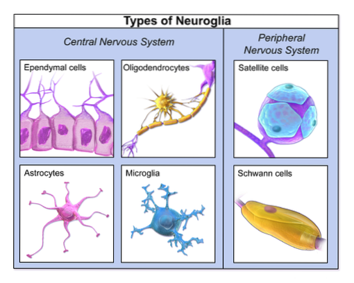
Together with oligodendrocytes, microglial cells, and ependymal cells, astrocytes form what is known as "neuroglia." The neuroglial cells are usually found in much greater numbers than neurons, but they do not participate in the reaction and / or propagation of nerve impulses..
The terms "neuroglia" and "astrocyte" were proposed in 1895 by Mihaly von Lenhossek to identify the cell group that supports neurons and a special class of these cells, characterized by their stellate shape..
Astrocytes have been shown to increase the number of functional neuronal synapses in neurons of the central nervous system, which means that they are required for the transmission of nerve stimuli..
These cells comprise between 20 and 25% (and sometimes up to 50%) of the volume in many brain areas and are known to have special roles in responding to injury, although it has recently been proposed that they are involved in many diseases of the system. central nervous.
Article index
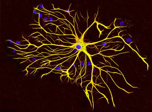
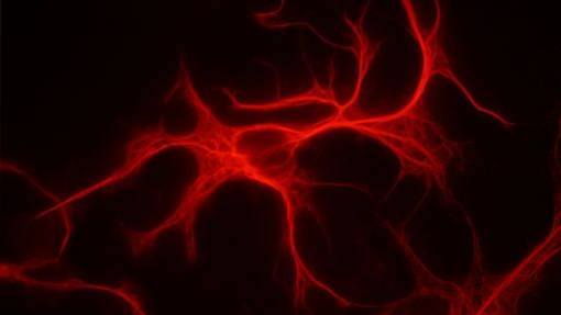
Astrocytes are "stellar" or star-shaped cells, as they have cytosolic projections of different sizes that make them similar to children's drawings of a space star.
These cells are distributed throughout the brain and throughout the spinal cord and constitute more than 50% of all glial cells..
When viewed under a light microscope after routine staining, astrocytes (depending on type) have large oval or lobulated nuclei with little cytosolic content..
The characteristic cytosolic projections of astrocytes are known as "glial fibrils", and are mostly composed of the glial-fibrillar acidic protein (GFAP). Glial Fibrillary Acidic Protein), specific for astrocytes of the central nervous system and is commonly used as a marker protein.
Glial fibers of astrocytes are closely related to the cell body and axons of neurons, surround the sites of nerve synapses and also the well-known nodules of Ranvier, present in axons covered by a myelin sheath.
Although they are not excitable cells, astrocytes express specific sodium and potassium channels that are very important for their functions in maintaining the homeostasis of the nervous system..
Astrocytes have two types of specializations in their membranes known as junctions gap and orthogonal assemblies.
Unions gap are made up of transmembrane proteins called connexons, which join with homologous proteins in nearby cells to form hydrophobic channels through which small molecules can exchange between cells.
There are numerous type unions gap between astrocyte-astrocyte and between astrocytes and oligodendrocytes. Among the molecules that are exchanged through these bonds are small ions, oligosaccharides and certain trophic factors..
Orthogonal assemblies, on the other hand, are "paracrystalline" arrangements that are made up of 7 nm particles. They are numerous in the more distal portions of the cytosolic projections, especially in the region facing the blood vessels.
These structures participate in cell adhesion and in the transport of substances between astrocytes and between astrocytes and the cerebrospinal fluid..
There are two well-defined types of astrocytes that differ in their morphology and anatomical location. These are protoplasmic astrocytes and fibrous astrocytes..
However, many researchers consider that they are the same type of cells that acquire different functions depending on the environment where they are..
Other bibliographic documents, however, establish the existence of a third type of astrocytes, characterized by their elongated cell bodies and commonly known as the glial Bergmann cells of the cerebellum and the Müller cells in the retina of the eyes..
Only the astrocytes present in the brain and spinal cord will be described here..
The existence of such cells was demonstrated by silver staining techniques. These are typical of the gray matter of the brain and are cells with a stellar aspect (similar to a star).
They have an abundant cytosol where a large nucleus is found and they differ from fibrous astrocytes in that they have short processes.
The ends of some of the cytosolic projections are composed of "vascular feet" or pedicels that interact with adjacent blood vessels..
Some protoplasmic astrocytes are close to the cell bodies of some neurons, as if they were "satellite" cells.
Fibrous astrocytes are cells with few internal organelles, rich in free ribosomes and storage molecules such as glycogen. They have longer cytosolic projections or projections than protoplasmic astrocytes, which is why they are known as “fibrous” astrocytes.
These cells are associated with the white matter of the brain and their processes also connect with blood vessels, but are separated from these by their own basal lamina.
As neuroglial cells, astrocytes play a vital role in the physical support and metabolic support of neurons of the central nervous system in vertebrate animals..
In addition, these cells are responsible for the elimination of ions and other waste substances of neuronal metabolism that are typical of the neuronal microenvironment, especially of the axonal region, such as, for example:
- Potassium Ions (K +)
- Glutamate residues and
- Gamma aminobutyric acid (GABA) residues
In charge of, among other things, the energy metabolism of the cerebral cortex, as they release glucose from glycogen molecules stored in their cytosol.
This release occurs only when astrocytes are stimulated by neurotransmitters such as norepinephrine and vasoactive intestinal peptide or VIP peptide, which are released by nearby neurons..
Astrocytes also participate in neuronal development and in the transport and release of neurotrophic factors, which is why some authors consider that they are cells that maintain homeostasis in the central nervous system.
These cells can also play important roles in healing damaged areas of the brain. They control brain pH and regulate multiple neural functions by maintaining a relatively constant microenvironment.
Some astrocytes participate in the formation and preservation of the blood-brain barrier, as they have the ability to form a continuous layer on the blood vessels in the periphery of the central nervous system..
The blood-brain barrier is a kind of "structure" that limits the entry of circulating blood elements into the central nervous system..
The relationship of these nerve cells with this function in such that it has been experimentally demonstrated that epithelial cells can induce the differentiation of astrocytic precursors.
Some bibliographic reviews highlight astrocytes as immunocompetent cells of the central nervous system, since they are capable of expressing proteins of the major histocompatibility complex class II (MHC). Major Histocompatibility Complex), which have important roles in antigen presentation.
These cells, then, participate in the activation of T cells, not only by the expression of antigen-presenting proteins, but also by their ability to express co-stimulatory molecules that are critical for the process. per se.
However, the participation of astrocytes in the immune system is not limited to the presentation of antigens, but it has also been shown that these cells can secrete a wide variety of cytokines and chemokines, which may mean that they are involved in inflammatory processes and immune reactivity in the brain.
In view of experimental data suggesting that the suppression of astrocytes in the central nervous system leads to substantial neuronal degeneration in adults, it is clear that these cells have valuable clinical significance..
Astrocytes, among their multiple functions, have been linked to the long-term recovery of patients with brain injuries. They are also involved in the regeneration of neurons, mainly due to their ability to express and release trophic factors..
In other words, the survival of neurons is highly dependent on their association with astrocytes, such that any massive damage that occurs in these cells will directly affect normal brain functions..
Many neurodegenerative diseases are distinguished by proliferation, morphological change, and increased expression of glial-fibrillar acidic protein (GFAP) in astrocytes; condition known as "astrogliosis".
This process, depending on the context where it occurs, can be beneficial or deleterious, as it can mean neuronal survival due to the production of growth factors or the formation of "glial scars", respectively..
Astrogliosis is not a random or "all or nothing" process. Rather, it is a highly controlled event that depends on multiple cellular signals and the particular context in which the cell in question is found..


Yet No Comments