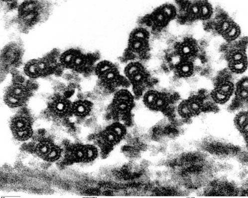
The axoneme It is an internal cytoskeletal structure of cilia and flagella based on microtubules and that gives movement to them. Its structure is made up of a plasma membrane that surrounds a pair of central microtubules and nine pairs of peripheral microtubules..
The axoneme is located outside the cell and is anchored inside the cell by means of the basal body. It is 0.2 μm in diameter and its length can vary from 5-10 μm in cilia to several mm in the flagellum of some species, although these generally measure 50-150 μm.
The axoneme structure of cilia and flagella is highly conservative in all eukaryotic organisms, from microalgae Chlamydomonas to the scourge of the human sperm.
Article index
The axonemes of the vast majority of cilia and flagella have a configuration known as "9 + 2", that is, nine pairs of peripheral microtubules surrounding a central pair.
The microtubules of each pair are different in size and composition, except for the central pair, which presents both microtubules similar. These tubules are stable structures capable of resisting ruptures..
Microtubules have polarity and all have the same arrangement, with their “+” end located towards the apex and the “-” end located basally..
As we already pointed out, the structure of the axoneme is of type 9 + 2. Microtubules are long cylindrical structures, made up of protofilaments. Protofilaments, in turn, are made up of protein subunits called alpha tubulin and beta tubulin..
Each protofilament has an alpha tubulin unit at one end, while the other end has a beta tubulin unit. The end with the beta tubulin terminal is called the "+" end, the other end would be the "-" end. All protofilaments of the same microtubule are oriented with the same polarity.
Microtubules contain, in addition to tubulins, proteins called microtubule-related proteins (MAPs). Of each pair of peripheral microtubules, the smallest (microtubule A) is made up of 13 protofilaments.
Microtubule B has only 10 protofilaments, but it is larger than microtubule A. The central pair of microtubules has the same size and each of them is made up of 13 protofilaments.
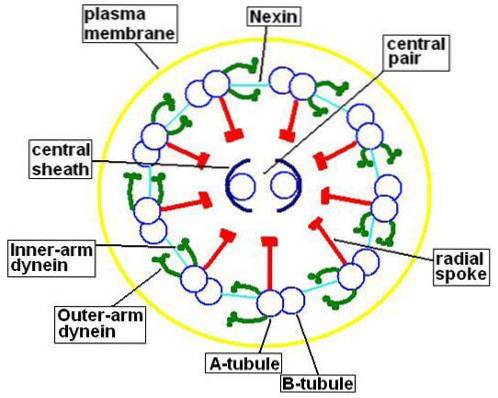
This central pair of microtubules is enclosed by the central sheath, protein in nature, which will connect with the peripheral A microtubules by means of the radial rays. On the other hand, the microtubules A and B of each pair are joined together by a protein called nexin..
Microtubules A part also a pair of arms formed by a protein called dynein. This protein is responsible for using the energy available in ATP to achieve the movement of cilia and flagella.
Externally, the axoneme is covered by a ciliary or flagellar membrane that has the same structure and composition as the plasma membrane of the cell..
Although the “9 + 2” composition of the axoneme is highly conserved in most eukaryotic ciliated and / or flagellated cells, there are some exceptions to this model..
In the sperm of some species, the central pair of microtubules is lost, resulting in a "9 + 0" configuration. The flagellar movement in these spermatozoa does not seem to vary much from that observed in axonemes with normal configuration, for which it is believed that these microtubules do not have an important participation in the movement..
This axoneme pattern has been observed in the sperm of species such as fish Lycondontis and of annelids of the genus Myzostomum.
Another configuration observed in axonemes is the “9 + 1” configuration. In this case, a single central microtubule is present, rather than a pair. In such cases, the central microtubule is extensively modified, presenting several concentric walls.
This axoneme pattern has been observed in the male gametes of some species of flatworms. In these species, however, this axoneme pattern is not repeated in other flagellated or ciliated cells of organisms..
Studies of flagella movement have shown that flagella flexion occurs without contraction or shortening of the microtubules of the axoneme. Due to this, the cytologist Peter Satir has proposed a model of flagellar movement based on the displacement of microtubules..
According to this model, movement is achieved thanks to the displacement of a microtubule from each pair on its partner. This pattern is similar to the sliding of myosin chains on actin during muscle contraction. Movement occurs in the presence of ATP.
The dynein arms are anchored in microtubule A of each pair, with the ends directed toward microtubule B. At the beginning of movement, the dynein arms adhere to the binding site on microtubule B. Then, a change occurs in the configuration of the dynein that drives microtubule B down.
Nexin keeps both microtubules close to each other. Subsequently, the dynein arms separate from microtubule B. It will then rejoin to repeat the process. This sliding occurs alternately between one side of the axoneme and the other..
This alternating displacement on one side of the axoneme causes the cilium, or flagellum, to bend first to one side and then to the opposite side. The advantage of Satir's flagellar movement model is that it would explain the movement of the appendix independently of the axoneme configuration of the axoneme microtubules.
There are several genetic mutations that can cause abnormal development of the axoneme. These abnormalities can be, among others, the lack of one of the dynein arms, either internal or external, of the central microtubules or of the radial rays..
In these cases, a syndrome called Kartagener syndrome develops, in which people who suffer from it are infertile because the sperm are not able to move.
These patients also develop viscera in an inverted position in relation to the normal position; for example, the heart located on the right side of the body and the liver on the left. This condition is known as situs inversus.
Those with Kartagener syndrome are also prone to respiratory and sinus infections.
Another disease related to abnormal development of the axoneme is polycystic kidney disease. In this, multiple cysts develop in the kidneys that end up destroying the kidney. This disease is due to a mutation in the genes that encode proteins called polycystins.


Yet No Comments