The bursa or synovial bag is an anatomical structure that owes its name to the fact that it has a shape very similar to that of a bag, with the particularity that it is hermetically sealed.
The bursa or bursa consists of a thin membrane that surrounds or surrounds a viscous and slippery liquid that it produces, and in turn forms the internal lining of the joint capsule. The internal fluid is called synovium or synovial fluid.
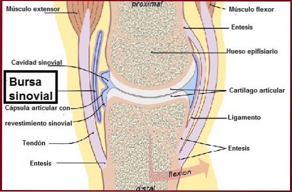
The synovial bag is a structure that performs a protective function of the anatomical joints where there is movement, that is, it prevents the bones from rubbing directly with other structures.
If the bag did not exist, the bones and other structures would wear down each other, and the movement of the joints would be almost impossible to perform due to the pain it would cause..
Therefore, as can be seen, the bursae are strategically placed between two anatomical structures in which there is sliding or movement; assuming the work of friction or rubbing.
The synovial fluid that contains the bursa is what keeps the walls of the bursa lubricated in its internal part; allowing the sliding between its walls.
The synovial bag must be kept intact to prevent synovial fluid from leaking or infiltrating. An involvement of the bursa produces an inflammatory clinical picture called bursitis, which can have various causes.
Article index
The name synovial bag comes from the Latin bursa, which means "bag". While, the word synovial comes from the Latin synovia composed of the Greek prefix syn- (with, together) and the Latin term ovum which means (egg), plus the suffix to the (relative to).
Then, according to the meaning of the words, it can be deduced that it is an airtight bag that contains inside a liquid similar to egg white in terms of appearance, color and texture..
Synovial bursae are present in joints of the diarthrosic type or also called synovials, differing from solid joints, where the bursa is absent..
The synovial bursae not only protect the junction between two bones, they are also present in other anatomical sites, that is, it separates a bone from a ligament, a tendon or simply from the skin. There are two types of bursa, deep and superficial.
This type of synovial bursa are those that protect from friction or friction between two bone structures or between a bone with nearby muscles or ligaments.
This type of synovial bursa, as its name indicates, is located more towards the surface and protects from friction or friction between a bone structure (bone or bone protrusion and the skin).
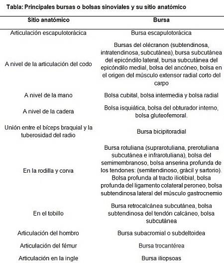
There are many synovial bursae distributed throughout the body. They are mainly found in joints with a lot of movement, or where the protection of certain structures is required. It is estimated that the human body can have up to a thousand synovial bags distributed throughout the locomotor system.
The names given to bursae are related to the anatomical site and the structure involved. The most relevant ones will be shown in the following table.
It is important to emphasize that the bursa and the structures that surround it (capsule and ligaments) receive blood vessels that feed them. It also receives sensory nerves that send information to the brain regarding the stress exerted on the joints..
This involvement is due to inflammation of the bursa or synovial bag. The bursa becomes inflamed for a variety of reasons, such as: overuse and repetitive use of a particular joint, infection, or trauma.
It can also be a consequence of previous diseases, such as rheumatoid arthritis, progressive systemic sclerosis, gout, among others..
The most frequent symptoms of bursa inflammation are: pain on palpation, limitation in movement of the affected joint and, very importantly, there is an increase in volume, because the bursa secretes more synovial fluid than normal, among others..
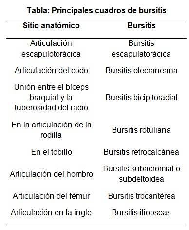
The names for bursitis depend on the anatomical site or joint involved. For example, inflammation of the bursa at the level of the scapulothoracic joint is called scapulothoracic bursitis, that of the elbow (olecranon bursitis) due to the olecranon bone.
While the inflammation of the bursa present between the biceps brachii and the tuberosity of the radius is called bicipitoradial bursitis, etc. See the following table.
Drinking alcohol increases the likelihood of post-traumatic infectious bursal disease. The same happens with the suffering of immunosuppressive diseases, since these patients are more likely to suffer from an infection at the joint level.
Patients suffering from an exaggerated elevation of uric acid or calcium can form crystals that accumulate in the joints and underlying tissues. These crystals damage and inflame the bursa.
On the other hand, students and people undergoing hemodialysis tend to place the elbow for hours on a very hard surface, so the continuous compression inflames the joint, causing olecranon bursitis..
Athletes are at higher risk of suffering from bursitis, as well as those who perform activities that require great physical effort (lifting heavy objects) or that involve repetitive movement.
Finally, people with autoimmune and degenerative diseases, such as osteoarthritis, arthritis, among others.
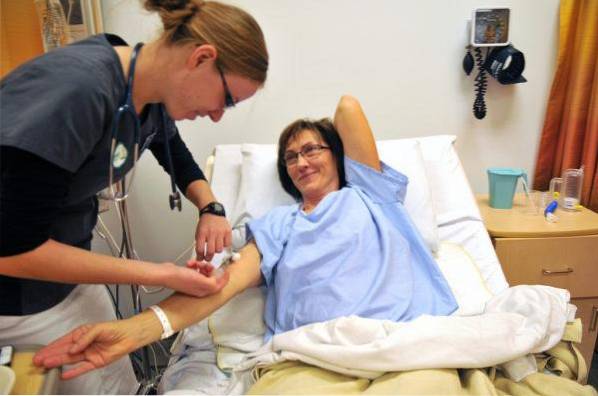
The presence of a fracture is ruled out by radiographic imaging and if it does not exist, then an ultrasound is performed. In case of suspected infectious bursal disease, a synovial fluid sample is required for microbiological analysis..
Rest, cryotherapy and elimination of the offending agent, either mechanical (a specific movement) or detoxification of elements that accumulate in the blood, such as taking medications to lower uric acid levels or finally the supply of antibiotics if the cause it is an infectious problem.
Massage is contraindicated in bursitis.
It is a rare, benign pathology, the cause of which is unknown. It is characterized by causing pain, inflammation and osteoarthritic changes at the level of the affected joint. The pain is especially accentuated after some physical effort.
Its diagnosis is made by observing radiologically inside the synovial fluid or in the joint capsule free bright white structures.
These fragments are of cartilaginous or osteocartilaginous origin, which are called "loose body", resembling a snow storm. They can also be found in tendons and ligaments.
The affected joints can be the following in order of frequency: knee, hip, elbow, wrist, ankle, with the least affected joints being the shoulder and the mandibular temporo. Usually only one joint is affected.
Although its cause is unknown there are theories of its origin.
In this particular, some authors think that these free bodies are pedicle nodules that have detached from the synovial membrane, to later float in the synovial fluid, these begin to grow and are later crushed into small pieces due to the movement of the joint itself.
Fragmented particles can grow back and the cycle repeats. The largest preserved fragments are those that have lodged in the synovial recesses.
Yet No Comments