The zygote It is defined as the cell that results from the fusion between two gametes, one female and the other male. According to the genetic load, the zygote is diploid, which means that it contains the complete genetic load of the species in question. This is because the gametes that originate it each contain half of the chromosomes of the species..
It is often known as an egg and structurally it is made up of two pronuclei, which come from the two gametes that originated it. Likewise, it is surrounded by the zona pellucida, which fulfills a triple function: to prevent any other sperm from entering, to keep the cells resulting from the first divisions of the zygote together, and to prevent implantation from occurring until the zygote reaches the site. ideal in utero.
The cytoplasm of the zygote, as well as the organelles that are contained in it, are of maternal origin, since they come from the ovum.
Article index
The zygote is classified according to two criteria: the amount of yolk and the organization of the yolk..
Depending on the amount of yolk the zygote has, this can be:
In general, the oligolecito zygote is one that contains very little yolk. Likewise, in most cases they are small in size and the nucleus has a central position..
A curious fact is that this type of egg originates, mainly, larvae that have free life.
The type of animals in which this type of zygote can be seen are echinoderms, such as sea urchins and starfish; some worms such as flatworms and nematodes; mollusks such as snails and octopuses; and mammals like humans.
This is a word made up of two words, "meso" which means medium, and "lecito" which means yolk. Therefore, this type of zygote is one that has a moderate amount of yolk. Similarly, it is located mainly in one of the poles of the zygote..
This type of egg is representative of some vertebrates such as amphibians, represented by frogs, toads and salamanders, among others..
The word polilecito is formed by the words “poli”, which means a lot or abundant, and “lecito”, which means yolk. In this sense, the polylecyte zygote is one that contains a large amount of yolk. In this type of zygote, the nucleus is located in a central position of the yolk.
The polycyte zygote is typical of birds, reptiles and some fish such as sharks.
According to the distribution and organization of the yolk, the zygote is classified into:
The word isolecito is made up of "iso", which means equal, and "lecito", which means yolk. In such a way that the isolecyte-type zygote is one in which the yolk presents a homogeneous distribution throughout the available space..
This type of zygote is typical of animals such as mammals and sea urchins.
In this type of zygote, the yolk is abundant and occupies almost all the available space. The cytoplasm is quite small and contains the nucleus.
This zygote is representative of species of fish, birds and reptiles.
As can be inferred from the name, in this type of egg the yolk is in a central position. Likewise, the nucleus is in the center of the yolk. This zygote is characterized by being oval in shape.
This type of zygote is typical of members of the arthropod group, such as arachnids and insects..
The zygote is the cell that forms immediately after the fertilization process occurs.
Fertilization is the process by which the male and female gametes unite. In humans, the female zygote is known as the ovum and the male zygote is called the sperm..
Similarly, fertilization is not a simple and straightforward process, but is made up of a series of stages, each one very important, namely:
When the sperm makes the first contact with the ovum, it does so in the so-called zona pellucida. This first contact has a transcendental importance, since it serves for each gamete to recognize the other, determining if they belong to the same species.
Likewise, during this stage, the sperm is capable of crossing a layer of cells that surround the ovum and that together are known as the corona radiata..
In order to pass through this layer of cells, the sperm secretes an enzymatic substance called hyaluronidase that helps it in the process. Another element that allows the sperm to penetrate this outer layer of the ovum is the frenzied movement of the tail..
Once the sperm has crossed the radiated crown, the sperm faces another obstacle in order to penetrate the ovum: the zona pellucida. This is nothing more than the outer layer that surrounds the egg. It is made up mainly of glycoproteins.
When the head of the sperm comes into contact with the zona pellucida, a reaction known as the acrosome reaction is triggered. This consists of the release, by the sperm, of enzymes that as a whole are known as spermiolysins. These enzymes are stored in a space in the sperm's head known as the acrosome..
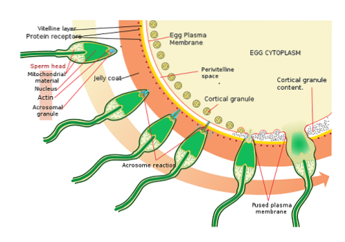
Spermiolysins are hydrolytic enzymes whose main function is the degradation of the zona pellucida, to finally fully penetrate the ovum.
When the acrosome reaction begins, a series of structural changes are also triggered in the sperm at the level of its membrane, which will allow it to fuse its membrane with that of the ovum..
The next step in the fertilization process is the fusion of the membranes of the two gametes, that is, the ovum and the sperm..
During this process, a series of transformations occur in the ovum that allow the entry of a sperm and prevent the entry of all the other sperm that surround it..
In the first place, a duct known as the fertilization cone is formed, through which the membranes of the sperm and ovum come into direct contact, which end up merging..
Simultaneously with this, a mobilization of ions such as calcium (Ca+two), hydrogen (H+) and sodium (Na+), which generates the so-called depolarization of the membrane. This means that the polarity that normally has is reversed.
Similarly, under the membrane of the ovule are structures called cortical granules, which release their content to the space that surrounds the ovule. With this, what is achieved is to prevent the adherence of the sperm to the ovum, so they will not be able to approach it..
For the zygote to finally form, it is necessary for the nuclei of the sperm and the egg to unite.
It is worth remembering that gametes contain only half the number of chromosomes of the species. In the case of the human being, it is 23 chromosomes; This is why the two nuclei must fuse to form a diploid cell, with the complete genetic load of the species.
Once the sperm enters the egg, the DNA it contains is duplicated, as well as the DNA of the pronucleus of the egg. Next, both pronuclei are next to each other.
Immediately, the membranes that separate the two disintegrate and in this way the chromosomes that were contained in each one can join with their homologue..
But everything does not end here. Chromosomes are located at the equatorial pole of the cell (zygote) to initiate the first of many mitotic divisions in the segmentation process..
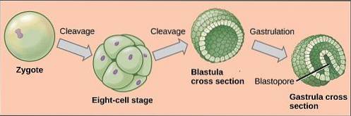
Once the zygote has formed, it begins to undergo a series of changes and transformations that consist of a successive series of mitoses that transform it into a mass of diploid cells known as a morula..
The developmental process that the zygote goes through includes several stages: cleavage, blastulation, gastrulation, and organogenesis. Each one of them is of preponderant importance, since they play a key role in the formation of the new being..
This is a process by which the zygote undergoes a large number of mitotic divisions, multiplying its number of cells. Each of the cells that form from these divisions is known as blastomeres..
The process occurs as follows: the zygote divides into two cells, in turn these two divide, originating four, these four into eight, these into 16 and finally these into 32.
The compact cell mass that forms is known as a morula. This name is because its appearance is similar to that of a blackberry.
Now, depending on the quantity and location of the yolk, there are four types of segmentation: holoblastic (total), which can be equal or unequal; and the meroblastic (partial), which can also be equal or unequal.
In this type of segmentation, the entire zygote is segmented through mitosis, resulting in blastomeres. Now, holoblastic segmentation can be of two types:
It is typical of zygotes that contain abundant yolk. In this type of segmentation, only the so-called animal pole is divided. The vegetative pole is not involved in the division, so that a large amount of yolk remains unsegmented. Likewise, this type of segmentation is classified as discoidal and superficial..
Here only the animal pole of the zygote experiences segmentation. The rest of this, which contains a lot of yolk, is not segmented. Likewise, a disc of blastomeres is formed that will later give rise to the embryo. This type of segmentation is typical of telolecyte zygotes, especially in birds and fish..
In superficial meroblastic cleavage, the nucleus undergoes various divisions, but the cytoplasm does not. In this way, several nuclei are obtained, which move towards the surface, being distributed throughout the cytoplasmic covering. Subsequently, the cellular limits appear that generate a blastoderm that is peripheral and that is surrounding the yolk that was not segmented. This type of segmentation is typical of arthropods.
It is the process that follows segmentation. During this process, the blastomeres attach to each other, forming very close and compact cell junctions. Through blastulation the blastula is formed. This is a hollow, ball-shaped structure with an internal cavity known as a blastocele..
It is the outer cell layer that is also called the trophoblast. It is of vital importance because from it the placenta and the umbilical cord will be formed, important structures through which an exchange between the mother and the fetus is established..
It is made up of a large number of cells that migrated from the interior of the morula to the periphery.
It is the internal cavity of the blastocyst. It is formed when the blastomeres migrate towards the external parts of the morula to form the blastoderm. The blastocele is occupied by a fluid.
It is an internal cell mass, which is located inside the blastocyst, specifically at one of its ends. From the embryoblast the embryo itself will be formed. The embryoblast in turn is made up of:
Both the epiblast and the hypoblast are extremely important structures, since from them the so-called germ leaves will develop which, after a series of transformations, will give rise to the various organs that make up the individual..
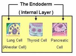
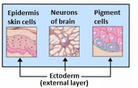
This is one of the most important processes that occur during embryonic development, since it allows the formation of the three germ layers: endoderm, mesoderm and ectoderm..
What happens during gastrulation is that the epiblast cells begin to proliferate until there are so many that they must move you the other way. In such a way that they move towards the hypoblast, managing to even displace some of its cells. This is how the so-called primitive line is formed.
Immediately, an invagination occurs, through which the cells of this primitive line are introduced in the direction of the blastocele. In this way a cavity known as the archenteron is formed, which has an opening, the blastopore.
This is how a bilaminar embryo is formed, made up of two layers: the endoderm and the ectoderm. However, not all living beings come from a bilaminar embryo, but there are others, such as humans, that come from a trilaminar embryo.
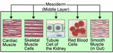
This trilaminar embryo is formed because the cells of the archenteron begin to proliferate and are even located between the ectoderm and the endoderm, giving rise to a third layer, the mesoderm..
From this germ layer the epithelium of the organs of the respiratory and digestive systems is formed, as well as other organs such as the pancreas and the liver..
It gives rise to bones, cartilage, and voluntary or striated muscles. Likewise, organs of the circulatory system and others such as the kidney, gonads and myocardium, among others, are formed from it..
It is responsible for the formation of the nervous system, skin, nails, glands (sweat and sebaceous), the adrenal medulla and the pituitary..
It is the process by which, from the germ layers and through a series of transformations, each and every one of the organs that will make up the new individual originate..
Broadly speaking, what happens here in organogenesis is that the stem cells that are part of the germ layers begin to express genes whose function is to determine what type of cell is going to originate.
Of course, depending on the evolutionary level of the living being, the organogenesis process will be more or less complex.


Yet No Comments