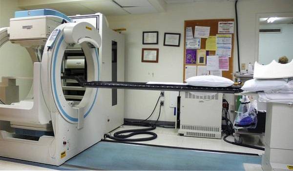
The bone scintigram It is a special imaging study that allows determining the presence of abnormalities in the skeleton. It uses nuclear medicine techniques and small amounts of radioactive contrast to “stain” the bones, which are later photographed using equipment very similar to those used to obtain X-rays..
This contrast - or more correctly, this tracer isotope - travels through the blood and is deposited in the bones. There it gives its radioactive capacity to bone tissue in the form of gamma rays, which are then detected by special sensors located in scintigraphy equipment. These devices produce an image similar to an X-ray.
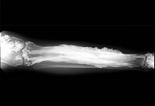
If there are alterations in the bones, whatever their cause, the uptake of the tracer isotope is modified. This change can be an increase in uptake (known as hyperuptake) or decrease (known as hypouptake). The results of these images are analyzed by a radiologist or other experienced specialist..
Article index
The bone scintigram has multiple applications in the medical world. Most of these are direct injuries to the bone or other systemic diseases that can affect the skeleton. The most important reasons for the indication of this study are infectious, oncological and traumatic processes..
This study has a very high sensitivity when there are alterations in bone metabolism. It can even detect early bone lesions when no significant clinical manifestations or apparent lesions on classical radiographs have even appeared.
One of the most frequent uses of the bone scintigraphy is the global visualization of the human skeleton. It is one of the few studies that allow this possibility, helping health professionals to evaluate all of the bones without the need to review several plates as is the case with traditional X-rays, CT scans or MRIs..
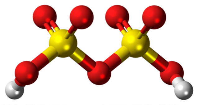
Osteomyelitis (infection of the bone) is the main indication for bone scintigraphy. Since the appearance of phosphates and polyphosphates, nuclear imaging has become essential in the diagnosis and control of bone infections, providing very valuable information to treating physicians..
The most requested exam is the three-phase scintigraphy. Depending on the time elapsed since the administration of the isotope, the perivascular space can be evaluated in a first phase, the bone fluid space in the second phase, and in the third and last phase the bone is evaluated as such..
Scintigraphy is the standard procedure for the detection of bone metastases generated by any primary tumor. It is much more sensitive when there are osteolytic lesions with a high osteoblastic response; this means that in lymphomas or several solid tumors, with poor osteoblastic response, it can give inaccurate information.
However, when used in conjunction with MRI, it is the ideal study to evaluate metastases..
It is also part of the usual protocol in primary bone tumors, although it is not the initial study since it does not allow the evaluation of the surrounding soft tissues or the necessary anatomical measurements..
There are many indications that the bone scintigraphy has in the trauma world. One of the advantages is its use in early and hidden lesions that, despite having clinical manifestations, cannot be detected with classical radiological studies. It is also combined with MRI for better results.
Sports injuries can be detected through this study. For a long time it was the quintessential imaging exam when stress fractures or medial tibial stress syndrome were suspected, but it has lately been superseded by the same MRI and its specialized variants.
In other medical conditions, a bone scan may be indicated. Among the most important we have the following:
Arthritis, plantar fasciitis, polymyositis, and Paget's disease.
Hyperparathyroidism, osteomalacia, and acromegaly.
Osteochondritis of the hip, bone infarcts due to sickle cell disease, osteoid osteoma.
As it is not a laboratory test, there is no range of values or levels considered normal. The evaluation of the results is based on the two conditions mentioned above: hyper-uptake or under-uptake of reactive tracer isotopes..
Most of the diseases that affect the bone produce hyper-uptake of the radioactive isotope used. This is due to the normal periosteal and osteoblastic inflammatory response that is generated in the bone tissue in the event of an attack, which is a factor that favors uptake..
The vast majority of oncological diseases that cause bone lesions or metastases - with the exception of lymphoma and some solid tumors - generate hyperuptake of the tracer. The same occurs with infectious processes, in which the image is conclusive for the diagnosis of osteomyelitis..
Traumatological injuries that generate a solution of continuity in the bone, especially if there is vascular damage, can cause local underuptake with periosteal or perivascular hyperuptake. It is evident that, in the absence of adequate blood supply, the isotope does not reach the site of injury.
Some benign tumors such as cysts or osteomas, as they are not vascularized, are hypocaptant lesions. This phenomenon also occurs when chronic injuries are not treated correctly and bone tissue becomes devitalized..
As there is no osteoblastic reaction or production of new bone, there is no uptake or subsequent generation of gamma rays.
To carry out this study, certain caution must be exercised in some cases, even with absolute contraindications..
Although extremely rare, allergic reactions to radiopharmaceuticals can occur. They are usually mild and do not generate complications, but they should not be ignored.
Isotope interactions have also been described with certain medications, some commonly used such as corticosteroids, nifedipine, and iron..
Administration of a tracer isotope can cause pain, phlebitis, and reddening of the skin. This reaction is highly dependent on the infusion rate and the caliber of the vessel into which the catheter was inserted. The discomfort disappears quickly and does not limit the performance of the study.
There is always some risk of injury and cell death when a radioactive isotope is administered, despite its low radiation level. It happened more frequently with phosphates, but today tracers are much safer.
The vast majority of authors recommend that the study be delayed until the end of pregnancy and breastfeeding..
If the clinical condition of the woman makes it necessary to carry out the study, she must be aware of the possible complications regarding her health and that of the fetus. Chances of miscarriage, stillbirth, and birth defects are high.

Yet No Comments