Greater circulation

2682
Robert Johnston
Greater circulation
The greater circulation is the one that makes the blood travel between the heart to the organs and tissues of the human body

What is the major circulation or systemic circulation?

The major circulation or systemic circulation is the circuit of our circulatory system through which blood moves from our heart to all the organs and tissues of our body.

The circulatory system is the organ system that is responsible for the movement of fluids in our body. Blood is one of those fluids, and perhaps the most important, since it carries very special components that our cells need to survive on a daily basis: oxygen and nutrients..

The cardiovascular system, which is the part of the circulatory system that is responsible for the movement of blood, is formed mainly by the heart and by all the blood vessels, which function as the system of tubes and channels through which the blood circulates throughout and width of our body.

Divisions of the circulatory system

The blood vessel complex of the circulatory system is "subdivided" into two circuits, one longer than the other:

  • Pulmonary circulation or less.
  • Pulmonary or systemic circulation.

Both circuits begin and end in the heart, so the entire system is a closed and practically continuous circuit.

The pulmonary circuit is the shorter of the two, since it comprises the movement of blood between the heart and the lungs: from the heart, blood without oxygen is pumped to the lungs, where it is oxygenated and sent back to the heart , and so on with each heartbeat.

The systemic circuit is longer, as it has to do with the movement of blood from the heart to all body tissues: those of the head and arms, those of the toes and legs, those of the visceral organs like the stomach, intestines, kidneys, liver, etc..

Route of the major circulation

The major circulation of our cardiovascular system begins and ends in the main organ: the heart..

How is the heart?

Human heart anatomy

To understand how this circuit works, it is necessary, first, to take into account some aspects of the structure of the heart, which, after all, is the one in charge of moving all the blood.

The heart is a hollow organ, formed by muscular walls that are capable of spontaneously contracting (without the help of the nervous system) to expel the blood inside and direct it towards its destination..

Our life depends on our heart contracting and relaxing continuously: when we sleep, when we eat, when we bathe, when we study and when we do any activity.

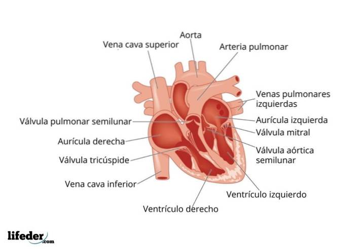
This organ is the size of a fist and we find it in the middle-left region of our chest, just between the lungs. Its shape is more or less conical and it is composed of 4 chambers connected to each other.

These chambers are known as the atria and ventricles. There is an atrium and a ventricle on the left side, and an atrium and another ventricle on the right side of the heart.

The atria are the upper chambers (also known as booster pumps) and the ventricles are the lower chambers, which are the real bombs that are responsible for the propulsion of the blood.

The atria connect with their respective ventricles, but the right region of the heart is not physically connected with the left region, that is, they do not exchange blood directly. These communicate through the pulmonary circulation.

The blood vessel system

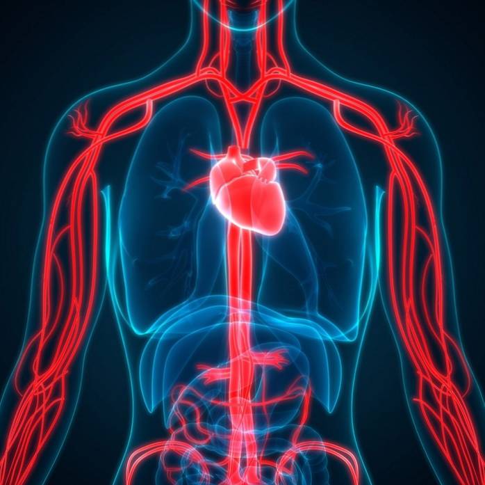
Blood moves between our heart and our body through the blood vessels that we know as arteries, veins, and capillaries..

  • The arteries They connect to the heart through the ventricles and therefore carry blood out of the heart. Its ramifications are known as arterioles.
  • The veins they connect to the heart through the atria and therefore carry blood back to the heart, either from the rest of the organs or from the lungs. The ramifications of the veins are known as venules.
  • The capillaries they are the smallest vessels and are the site where all the exchange of gases and nutrients occurs between the blood and the tissues.

How is the route of the major or systemic circulation?

Diagram of the major circulation

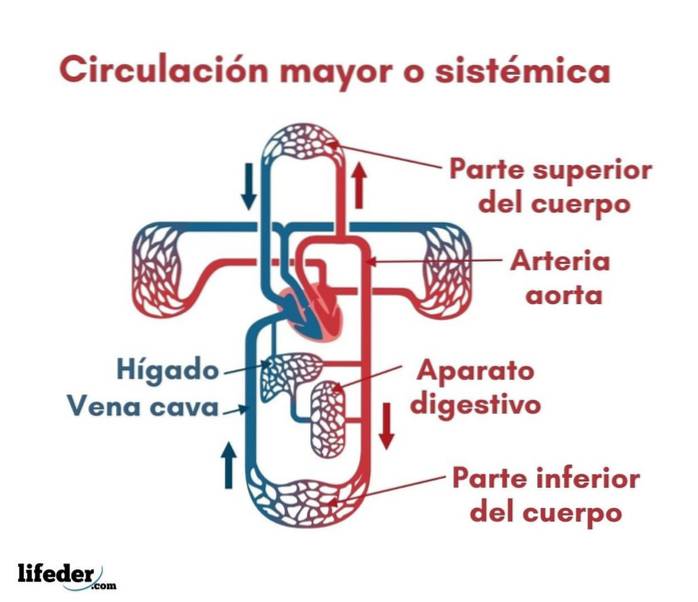
We can analyze the systemic circuit from the heart. Quickly it is as follows:

  1. Oxygenated blood leaves the heart and into the body tissues.
  2. Through the arteries, arterioles and capillaries, this blood is delivered to the organs and tissues, where gas exchange occurs: the blood delivers oxygen and nutrients and receives metabolic waste such as carbon dioxide and other potentially toxic molecules.
  3. Veins and venules carry blood back to the heart.
  4. Once there, the blood -deoxygenated- passes to the pulmonary circuit for oxygenation and when it reaches the heart again it is sent back to the rest of the body.
  5. This repeats with every heartbeat.

Let's see the route in more detail:

  1. Blood that has been oxygenated in the pulmonary circuit enters the left atrium from the lungs through the pulmonary veins.
  2. Once in the left atrium, it is propelled into the left ventricle, from where it is pumped into the body through the artery. aorta.
  3. From the aorta, the blood is transported by the arterial branches, by the arterioles and the capillaries, where oxygen and nutrients are delivered to the tissues, this is the systemic blood.
  4. When systemic blood is loaded with waste (carbon dioxide and others), this is drained or collected by the venules and transported to the veins to return to the heart.
  5. So the blood deoxygenated enters the right atrium of the heart through the vena cava.
  6. From the right atrium, blood is sent to the right ventricle and from there it leaves the heart through the pulmonary arteries to be oxygenated in the pulmonary circuit.
  7. Blood flow is maintained thanks to the continuous contraction and relaxation of the heart, which are permanently pushing the blood towards both circuits.

Themes of interest

Pulmonary circulation or less

References

  1. Berne, R., & Levy, M. (1990). Physiology. Mosby; International Ed.
  2. Gartner, L., & Hiatt, J. (2002). Text Atlas of Histology (2nd ed.). México D.F .: McGraw-Hill Interamericana Editores.
  3. Putz, R., & Pabst, R. (2006). Sobotta-Atlas of Human Anatomy: Head, Neck, Upper Limb, Thorax, Abdomen, Pelvis, Lower Limb; Two-volume set.
  4. Weinhaus, A. J., & Roberts, K. P. (2005). Anatomy of the human heart. In Handbook of Cardiac Anatomy, Physiology, and Devices (2nd ed., Pp. 59-85). Humana Press Inc.
  5. West, J. B. (1991). Physiological basis of medical practice. Williams & Wilkins.

Yet No Comments