
Clostridium difficile It is a gram positive bacterium that belongs to the group of firmicutes and is also part of the bacterial flora of the intestine. It was isolated in 1935 by Hall and O'Toole.
It constitutes a bacterium of a pathogenic type, specifically at the intestinal level. Infections with these bacteria are very common in people who have been on a long-term antibiotic regimen..

This is a bacterium that in recent years has become a real problem, especially in hospitals, as the number of patients infected with it increases more and more. In addition, to this is added the high resistance it has to common hygiene measures.
Some specialists consider that perhaps this resistance is due to the development of a strain that has mutated, has acquired resistance to conventional drugs and is more virulent.
The age group most vulnerable to infection by Clostridium difficile it is the elderly, who by nature have an immune system more prone to depression. This has been demonstrated by the numerous statistics that accompany the various studies that have been conducted on the subject..
The Clostridium difficile is a bacterium that if not treated in time can cause serious complications, including death.
Article index
The taxonomic classification of the Clostridium difficile is the next:
Domain: Bacterium
Division: Firmicutes
Class: Clostridia
Order: Clostridial
Family: Clostridiaceae
Gender: Clostridium
Species: Clostridium difficile
The Clostridium difficile It is a bacterium that is rod-shaped (elongated). They have rounded edges and flagella on their surface. They measure 0.5-3 microns wide by 6 microns long.
Cells are surrounded by a cell wall that is made up of a thick layer of peptidoglycan. It also has polymers, known as PSI, PSII and PSIII.
These polymers are similar to teichoic acid and lipoteichoic acid, present in other gram positive bacteria. The components of the cell membrane have been the object of study because they have an indispensable role in the therapeutic area.
In the cultures, slightly elevated, translucent colonies are observed, which have a crystalline mottling. In the same way, they give off a characteristic manure smell.
The DNA of this bacterium is concentrated in a circular chromosome, which has 29% nucleotides of cytosine and guanine. Likewise, it presents a circular plasmid that contains 28% nucleotides of the same type mentioned..
The Clostridium difficile it turns purple when subjected to Gram stain. This indicates that its cell wall contains peptidoglycan, which, due to its structure, retains the dye molecules, causing it to adopt the aforementioned color..
This bacterium produces spores when environmental conditions are unfavorable. These spores can survive for a period of about two years in harsh conditions. Once these change and become favorable, the spores germinate creating new cells of the bacteria.
The Clostridium difficile It has a metabolism that is based mainly on the fermentation of some sugars, among which the main one is glucose. Likewise, it also ferments fructose, mannitol, mannose and cellobiose..
This bacterium is ubiquitous. It is present in the normal microbiota of the human gastrointestinal tract as a commensal. It is also found in soil, sand, and hay. It has also been isolated from farm animals, rodents, and domestic animals such as cats and dogs..
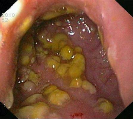
The Clostridium difficile it is considered a pathogenic agent, since through spores it is capable of generating certain pathologies. It has a preference for the gastrointestinal tract, where it germinates and causes diseases such as pseudomembranous colitis.
This bacterium can thrive under a variety of growth conditions. The accepted temperature range is between 25 and 45 ° C. Its optimum temperature is 30-37 ° C.
The bacterium produces two toxins, A and B. Both toxins act at the level of the epithelial cells of the intestine, triggering a series of changes that lead to the development of pathologies such as Diarrhea Associated with Clostridium difficile, Pseudomembranous Colitis and Antibiotic Associated Diarrhea.
This bacterium is not capable of synthesizing the enzyme catalase. This means that it cannot unfold hydrogen peroxide (HtwoORtwo) in water and oxygen.
The Clostridium difficile synthesizes gelatinase enzymes, which allow it to cause gelatin to liquefy. This is evident in the cultures, in which a transparent halo is observed around the colonies..
This bacterium does not synthesize the group of enzymes known as tryptophanases. Because of this, it is not capable of breaking the indole out of the tryptophan amino acid molecule. This is a test that allows differentiating the Clostridium difficile of other bacteria and even of others of the genus Clostridium.
The bacteria are capable of hydrolyzing urea to carbon dioxide and ammonia. This is because it does not synthesize the enzyme urease, since it does not have the genes for this..
The Clostridium difficile it does not synthesize the enzyme nitrate reductase therefore it cannot reduce nitrates to nitrites. This also constitutes a test for the identification and differentiation of bacteria..
This bacterium is a recognized human pathogen. It causes some diseases such as pseudomembranous colitis. The bacteria enter the body orally, mainly through contact with infected people.
The course of infection depends on whether the vegetative forms or the spores are ingested. In the first case, the living forms of the bacteria are eliminated in the stomach, thanks to the high level of acidity there..
Rather, spores are designed to withstand harsh environmental conditions, thus effectively resisting stomach conditions..
The spores manage to reach the small intestine and germinate there, thus producing the vegetative forms of the bacteria. These reach the large intestine where conditions are ideal for it to reproduce. Here it colonizes the mucosa, causing the presentation of the symptoms that characterize pseudomembranous colitis..
This disease can also be caused through another mechanism. When people are subjected to prolonged antibiotic therapy, this causes the gastrointestinal microbiota to suffer an imbalance.
This generates that the Clostridium difficile, which is a regular inhabitant of this flora, proliferates uncontrollably, giving way to disease.
The virulence factors that contribute to the bacterium Clostridium difficile causing damage to the gastrointestinal mucosa are the following:
Among the most prominent symptoms of intestinal pathology caused by Clostridium difficile can be mentioned:
At the level of the intestinal epithelium, certain lesions can be seen that indicate the evolution of the disease:
When it is suspected that a person may be showing signs and symptoms of an infection by Clostridium difficile, certain tests are carried out to reliably diagnose it.
Among these tests are the following:
When the clinical picture is caused by the previous administration of antibiotics, the first measure is to suspend said medication. It is expected that with this measure the table will be reversed.
If this does not happen, it is decided to administer an antibiotic treatment with drugs to which the bacteria are notably susceptible. Among these, the most recognized and used are metronidazole and vancomycin..


Yet No Comments