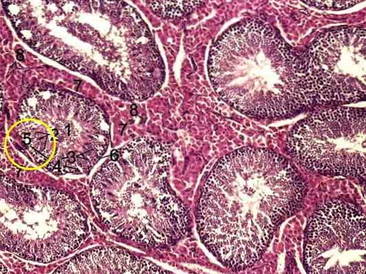
The Sertoli cells They are a type of sustainable cells located in the walls of the seminiferous tubes of the testes that participate in spermatogenesis. Sustainability cells are cells whose main function is to provide structural support in tissues and organs..
They are cells much taller than they are wide, with a large, irregular nucleus displaced towards the base of the cell. Their formation is controlled by the SRY gene and their number remains constant throughout the life of the organism, that is, they do not present mitotic divisions..
The functions of Sertoli cells include regulating the development and early stages of Leydig cell functioning, phagocytosis of residual cytoplasm during spermatogenesis, producing different hormones, and shaping the hermatotesticular barrier..
Among the diseases associated with Sertoli cells are the Sertoli-Leydig cell tumor and the Sertoli cell syndrome or germinal aplasia.
Article index
Sertoli cells were discovered by the Italian physiologist Enrique Sertoli in 1865. Sertoli who worked with various topics in human physiology, including the mechanisms of smooth muscle contraction, tissue carbonic acid and cellular proteins, discovered these cells by studying physiology. testicular.
They were named as Sertoli cells for the first time by the Viennese histologist von Ebner, twenty years after their discovery. Until the middle of the last century, these cells received little attention, as evidenced by the fact that only about 25 works related to them had been published to date..
However, with the invention of the electron microscope and the development of new study techniques in biochemistry and molecular biology, interest in Sertoli cells increased exponentially, with about 500 investigations a year currently.
Sertoli cells are columnar cells much taller than they are wide, displaying branching cytoplasmic processes to support developing germ cells. The highest concentration of cell organelles is distributed towards the basal portion of the cell.
The cell nucleus is large and euchromatic, its shape changes throughout the seminiferous epithelium cycle, occasionally presenting deep invaginations of the nuclear membrane. Its location is generally close to the base of the cell, however, it can occasionally move towards the lumen of the seminiferous tube..
The nucleolus is also very large and is intensely stained with vital dyes. Generally, this nucleolus has three clearly differentiable regions, that is, it is tripartite..
The total number of Sertoli cells will determine the maximum amount of sperm that a testicle can produce. The total volume of these cells in an individual is highly variable depending on the species, with a range that goes from 2000 to 7000 μm³.
However, there appears to be an inverse relationship between total volume and spermatogenic efficiency. These tubular cells extend from the basement membrane into the seminiferous epithelium lumen and have a “nurse-like” function on developing germ cells..
To perform this function, Sertoli cells extend their cytoplasm in projections in the form of thin arms and a cylindrical process which surround the spermatids and form complex specialized junctions that work as gap and tight junctions. They also use actin filaments and smooth endoplasmic reticulum.
The nucleus of the Sertoli cell is located, in most species, close to the basement membrane. It is large, elongated and occasionally its shape and location can be altered depending on the stage of the seminiferous cycle..
In the adult, the nucleus has deep invaginations of its membrane that give it an irregular shape and is surrounded by intermediate vimentin filaments. Additionally, it has a high density of pores in its membrane. Some proteins can occur in high concentrations near areas of intussusception..
The nucleolus is large and in many species it is made up of three easily distinguishable parts. It has from one to ten chrome centers.
The cytoplasm has numerous organelles that are arranged in a polarized way, that is, there is a higher concentration of organelles towards the basal portion of the cell than towards the distal portion.
Mitochondria are very abundant and can be elongated (2-3 μm), they can be cup-shaped or even donut-shaped. The rough endoplasmic reticulum is present in the basal area of the cell, while the smooth endoplastatic reticulum is the most abundant organelle in Sartoli cells..
Microtubules help maintain the distribution of the endoplasmic reticulum, as well as keep the mitochondria aligned. Sartoli cells possess phagocytic activity, for which they possess numerous lysosomes and multivesicular bodies. The Golgi apparatus, meanwhile, is relatively small.
Sertoli cells have been described as stem cells or nurse cells. One of the nursing activities they carry out is linked to the transport of iron, micronutrients and other substances to the developing germ cell by means of proteins such as transferrin and ceruloplasmin.
In addition to providing iron required for germ cell development, Sertoli cells also remove and recycle potentially toxic iron from residual bodies. Some authors call this last function as recycling and waste material management.
The secretory function of Sertoli cells is represented by hormones that can have autocrine, paracrine, and even endocrine activity. Paracrine functions include, for example, the signaling of germ cells to be targeted by follicle-stimulating hormones and tetosterone..
Additionally, after reaching puberty, Sertoli cells can regulate the production of follicle-stimulating hormone through the secretion of inhibin and activin, which act together..
It also produces various growth factors with paracrine activity, such as insulin-like growth factor 1 (IGF1), fibroblast growth factor (FGF), as well as transforming alpha (TGFA), which regulate the transformation of peritubular cells into Leydig cells, in addition to regulating the functioning of these.
Other hormones secreted by Sertoli cells that act during sex cell production include androgen-binding protein (ABP), estradiol, and glial cell-derived neutrophic factor (GDNF)..
Sertoli cells provide the testes with a unique immunoregulatory status, which has been demonstrated by transplanting testicular tissue into other different tissues, managing to survive for prolonged periods of time..
This is because, otherwise, the meiotic condition of the sex cells could cause them to be recognized by the antibodies as exogenous and potentially pathogenic factors and consequently activate the defense mechanisms for their destruction..
Among the molecules produced and secreted by Sertoli cells with immunoregulatory activity are, for example, the FAS / FAS Ligand system, protease inhibitor 9, CD40, CD59 or TGF-beta.
In addition to the immunoregulatory activity of the Sertoli cells, which protect the germ cells, the occluding junctions between the germ cells create a barrier that physically isolates the compartments where lymphocyte spermatogenesis takes place..
This barrier forms during puberty, when sperm production begins, and a break in it can trigger an immune response and cause male infertility..
This barrier acts dynamically allowing the migration of spermatocytes from the basal to the adluminal areas of the spermatic tube, but preventing, as already mentioned, the passage of lymphocytes..
There are some diseases related to Sertoli cells, among which the following can be mentioned:
This type of tumor is rare, representing less than 1% of testicular tumors. It can present in three histological varieties:
Although on a few occasions (10-20%) it can become malignant, in cases where it can metastasize to lymph nodes, bones and lung, survival rates are low.
This type of tumor does not present a hereditary component and is not related to any syndrome. The average age at which it manifests is 45 years.
It is much more aggressive than the classic tumor and, unlike this one, it can be associated with inheritance or with various syndromes, such as Peutz-Jeghers, Bourneville and also Carney complex.
The evil can appear early (17 years) or late (40 years), in both cases being a different behavior on their part. In the first case it can present multifocality, bilaterality, as well as hormonal activity, while in the second case it does not. On the other hand, its aggressiveness is greater in cases of late onset.
It is the least aggressive of the three varieties and to date no case of malignant behavior has been described. The mean age of onset is 35 years and, as in the case of the late calcifying cell tumor, it does not present multifocality, bilaterality or hormonal activity.
Also known as germ aplasia, it is a syndrome characterized by infertility caused by non-obstructive azoospermia (absence of germ cells). The causes of the syndrome are varied and among them are genetic disorders, mainly Klinefelter syndrome.
Other causes that have been associated with this syndrome include a history of cryptorchidism and / or varicocele. However, a high percentage of cases are of unknown origin.
Also known as arrenoblastoma, it is a type of sex cord tumor that can cause ovarian or testicular cancer. Its greatest occurrence occurs in young adults. It is generally benign and slow to develop.


Yet No Comments