
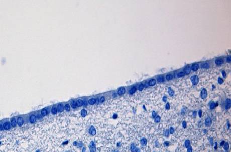
The ependymal cells, Also known as ependymocytes, they are a type of epithelial cell. They are part of the group of neurogliagles cells of the nervous tissue and line the cerebral ventricles and the central canal of the spinal cord..
This type of cell is characterized by presenting a cylindrical or cuboid shape and containing, in its cytoplasm, a large number of mitochondria and intermediate filamentous bundles..
At present, three main types of ependymal cells have been described: ependymocytes, tanicytes, and choroidal epithelial cells. Regarding their functionality, this type of cells seem to play an especially important role in the generation of cerebrospinal fluid and other substances..
Article index
Ependymal cells are a type of cell that is part of the neuroglia of nervous tissue. Thus, they are included within the set of neuroglial cells.
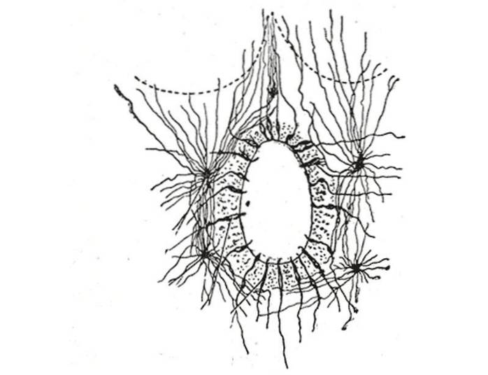
These cells stand out for forming the lining of the ventricles of the brain and the ependymal duct of the spinal cord. They have a columnar morphology and form a single layer of cubic and cylindrical cells..
Inside they have microvilli and cilia. These cilia are usually mobile, a fact that contributes to the flow of cerebrospinal fluid. Specifically, the cilia allow the fluid found on the cell surface to orient itself towards the ventricle..
The base of the ependymal cells lies on the inner glial limiting membrane. With regard to its cytoplasm, it is composed of mitochondria and intermediate filamentous bundles.
Finally, it should be noted that at the level of the cerebral ventricles, the ependymal cells undergo modifications. These modifications give rise to the formation of the choroid plexuses, vascular structures of the brain that are responsible for forming the cerebrospinal fluid..
Ependymal cells are formed from the embryonic neruoepithelium of the developing nervous system.
During the embryonic phase, the processes that arise from the cell body reach the surface of the brain. However, in adulthood, these extensions are characterized by being reduced and presenting only close terminations.
Through their development, ependymal cells generate, inside, a cytoplasm very rich in mitochondria and intermediate filamentous bundles.
Likewise, in their development process these cells acquire a ciliated shape in certain regions. These characteristics facilitate the movement of cerebrospinal fluid.
In brain structures where neural tissue is thin, ependymal cells form an inner limiting membrane that lines the ventricle and an outer limiting membrane just below the pia mater.
Finally, at the level of the cerebral ventricles, this type of cells are characterized by undergoing modifications and originating the choroid plexuses.
At present, three main types of ependymal cells have been described. This classification is carried out mainly through the encephalic location of each of them..
In this sense, ependymal cells can be divided into: ependymocytes, tanicytes and choroidal epithelial cells..
Ependymocytes are the most prevalent type of ependymal cells. Line the ventricles of the brain and the central canal of the spinal cord.
These types of cells are characterized by being in direct contact with the cerebrospinal fluid. Adjacent surfaces of ependymocytes possess junctions.
However, the cerebrospinal fluid communicates completely freely with the intercellular spaces of the central nervous system..
Tanicytes are the type of ependymal cells that line the floor of the third ventricle. Specifically, these cells are just above the median eminence of the hypothalamus.
They are characterized by having long basal processes that cross the cells of the median eminence. Likewise, they locate their terminal basal cells just above the blood capillaries..
The role of tanicytes is not well documented at present, although it has been attributed an important role in the transport of substances between the third ventricle and the hypothalamic median eminence..
Finally, the choroidal epithelial cells are the ependymal cells that are located in the cerebral ventricles. These cells are characterized by undergoing modifications and forming the choroid plexuses.
Both its base and its lateral regions form a series of folds. Epithelial cells are characterized by being held together through the tight junctions that surround them on their luminal surface..
The tight junctions between these cells are of vital importance in preventing the leakage of cerebrospinal fluid into the underlying tissues, as well as in limiting the entry of other substances into the cerebrospinal fluid duct.
The functions of ependymal cells are based mainly on the formation and distribution of cerebrospinal fluid..
Cerebrospinal fluid is a colorless substance that bathes both the brain and the spinal cord. It circulates through the subarachnoid space and the cerebral ventricles and is a basic substance to protect the brain.
More specifically, cerebrospinal fluid acts as a buffer to protect the central nervous system from trauma, provides nutritional elements to the brain and is responsible for eliminating metabolites
With regard to ependymal cells, their main functions are:
-They contain inside the cerebrospinal fluid that is produced in the choroid plexus, which is why they are vital cells when it comes to guaranteeing the protection of the central nervous system..
-Choroidal epithelial cells are responsible for directly producing cerebrospinal fluid. This fluid is secreted in the choroid plexuses, so without the functioning of this type of ependymal cells, the brain would lack cerebrospinal fluid..
-Certain studies postulate that ependymal cells also perform absorption functions since the free surfaces of ependymocytes present microvilli.
-Tanicytes are responsible for transporting chemicals from the cerebrospinal fluid to the pituitary portal system.
-At present it is postulated that ependymal cells could play a role in the control of hormonal production of the anterior lobe of the pituitary.


Yet No Comments