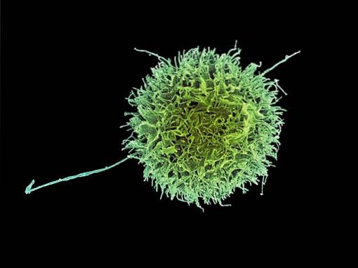
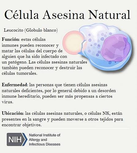
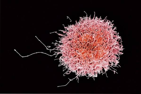
The NK cells (of English Natural Killer cells), natural killer cells or natural cytocidal cells, are a type of effector lymphocyte that participates in the responses of the innate or nonspecific immune system.
These cells were discovered more than 40 years ago and some authors describe them as “granular lymphocytes” that, unlike T and B lymphocytes, participate in the innate immune response and do not undergo genetic rearrangement processes in their germ lines..
Since they do not express the common markers for the other two classes of lymphocytes, NK cells were initially called "null cells." However, further studies showed that they were lymphocytes with large granulocytes..
These cells are capable of controlling different types of tumors and microbial infections by limiting their spread and tissue damage. In addition, they can lyse different types of cells without a defined antigenic stimulation..
NK cells are extremely important cells in the first line of defense against pathogens, a fact that has been shown through studies in which NK cell deficient humans can suffer lethal infections during childhood.
Article index
NK cells are found in a lower proportion than either of the other two classes of lymphocytes (they constitute 2 to 10% of circulating lymphocytes) and, since they belong to the innate defense system, it is thought that they were among the first cellular elements involved. in the protection of multicellular organisms.
Like T lymphocytes and B lymphocytes, NK cells are part of the mammalian hematopoietic system and are derived from progenitor hematopoietic cells that express CD34 + membrane markers, which are also known as HPC cells..
While T cells are known to mature in the thymus and B cells to mature in the bone marrow, attempts to determine the complete developmental pathway of NKs from HPC precursors have not been entirely successful; they are only known to be thymus-independent.
NK cells express adhesion molecules on their membrane surface known as CD2, LFA-1, NCAM, or CD56. They also express low-affinity receptors to the constant portion (Fc) of immunoglobulin IgG that are collectively called FcγRIIIA or CD16..
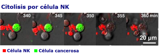
The interior of a natural cytocidal cell is packed with large cytosolic granules that are loaded with perforin, granzymes, and proteoglycans..
Perforins are pore-forming proteins that "pierce" the plasma membrane of cells that are attacked by NKs. Granzymes, on the other hand, are serine proteases that make their way into cells through the pores formed by perforins and degrade intracellular proteins..
The combined action of perforins and granzymes results in the arrest of the production of viral or bacterial proteins and in apoptosis or programmed cell death of the infected cell.
Natural killer cells function to eliminate “target” or “target” cells naturally, that is, spontaneously and without much specificity, since they do not require any type of antigenic priming..
One of the most important functions of this group of cells is its ability to kill tumor cells, especially those belonging to hematopoietic lineages, as well as cells invaded by different types of viruses and / or bacteria..
Its activity is strongly stimulated by factors such as IFN-α and β interferons, as well as by interleukin IL-12..
Thanks to the fact that these cells produce some important cytokines for the immune system, NKs participate in immune regulation, both in the innate and adaptive or specific systems..
For example, the production of interferon gamma (IFN-γ) in NK cells can disturb the participation of macrophages in innate immunity, as this molecule interferes with phagocytic and microbicidal activities.
At the same time, IFN-γ produced by natural cytocides can modify the commitment of entire populations of helper T cells, since IFN-γ also inhibits the expansion and development of one population relative to another..
NK cells represent the first line of defense during viral infections, as they control virus replication while cytotoxic T cells are activated, proliferate and differentiate, which can take more than 6 days.
NK cell populations are quite heterogeneous, both phenotypically, functionally and anatomically. In addition, its characteristics depend on the type of organism being studied..
In the murine (mouse) model, three different sets of natural cytocidal cells have been described that differ from each other by the expression of the markers CD11b and CD27. In this sense, there are the cells CD11bdullCD27 +, the CD11b + CD27 + and the CD11b + CD27dull.
The superscript "dull" refers to "off" or "inactive" and is used, in this case, to describe its state on the surface of murine cells..
CD11bdullCD27 + cells differentiate from a double positive type precursor (CD11b + CD27 +) which, in turn, gives rise to the more mature type of NK cells in rodents: CD11b + CD27dull.
Both the double positive lines and the CD11b + CD27dull lines are characterized by eliminating their target cells and by secreting a cytokine known as interferon (INF-γ). However, the latter are in something called "replicative senescence".
The three types of NK cells are distributed in different tissues. CD11bdullCD27 + cells are predominantly in lymph nodes and bone marrow. CD11b + CD27dull cells are abundant in the blood, spleen, lungs, and liver; meanwhile double positive cells have a more homogeneous or systemic distribution.
NK cells in humans are also classified according to the surface markers they express, but in this case they are differentiated by the presence of the markers CD56dim and CD56bright. The superscripts "dim" and "bright" refer to "dark" and "light", respectively..
The differences between these cells lie in the “target search” properties of each one, which are given by the presence of one or the other marker..
In the peripheral blood and spleen of humans the main type of NK cell is known as CD56dimCD16 +, which usually expresses the porphyrin protein and is cytotoxic. They also produce IFN-γ as a result of interaction with tumor cells under conditions in vitro.
CD56brightCD16- cells are found in lymph nodes and tonsils, which, instead of producing porphyrin, secrete the cytokine IFN-γ in response to stimulation by interleukins IL-12, IL-15 and IL-18.
In humans and rodents, the tonsils and other secondary lymphoid organs are thought to be the sites of production and maturation of most NK cells..
Some studies suggest that there is some similarity between human CD56bright cells and rodent CD11dull cells in terms of anatomical location, phenotypic characteristics, cytosolic perforin content, proliferative potential, and surface expression of interleukin IL-7R..
These have a fairly short half-life (approximately 2 weeks) and it is believed that in an adult human being there are around 2 trillion cells in circulation. They are abundant in the blood, spleen, and other lymphoid and non-lymphoid tissues.
Studies show that the normal concentration in adult men and women is around 200 and 600 cells per microliter of blood tested..
The intensity and quality of the cytotoxic responses of NK cells depends on the microenvironment generated by the cytokines and on the interaction with other cells of the immune system, especially with T cells, dendritic cells and macrophages..
Among the activating cytokines of NK cells are interleukins, specifically IL-12, IL-18 and IL-15; as well as type I interferon (IFN-I). Interferon and interleukins are potent activators of the effector function of NK.
Interleukin IL-2 is also involved in promoting proliferation, cytotoxicity, and cytokine secretion by NK cells..
IL-15 is crucial for the differentiation of NKs, while IL-2 and IL-18 are essential for the subsequent maturation of such cells..
Natural cytocidal cells are activated thanks to the recognition of their own molecules (a process known in English as “recognition of self molecules”) That are constitutively expressed under steady state conditions.
In their membranes, these cells express different members of a family of surface proteins that contain two or three immunoglobulin-like domains in their extracellular portions and motifs similar to the activation domains of immunoreceptors via tyrosine in their intracellular region..
Each NK cell can express one or more of these receptor proteins and each receptor is capable of recognizing a specific form of a major histocompatibility complex class I (MHC-I) molecule..
The recognition between this molecule and the receptor on the surface of natural cytocidal cells leads to the formation of a complex with abundant peptides derived from "self" proteins..
The receptors are mostly inhibitory proteins that activate a tyrosine phosphatase that prevents the cell from emitting normal responses.
The elimination or death mediated by natural killer cells is similar to that which occurs during the cytolytic action of CD8 T lymphocytes (cytotoxic), although the difference is that NKs are constitutive cytotoxic, that is, they do not need to be activated before.
Active NKs express the FasL ligand, which is why they relatively easily induce the death of target cells that express the Fas protein on their surface..
After the formation of the complete FasL / Fas, a process known as "degranulation" occurs, which ends with the release of porphyrin and granzymes at the intercellular contact sites..
Despite the aforementioned similarities, NKs differ from cytotoxic T-cell-mediated mechanisms in that the recognition of their target cells is not dependent on proteins of the major histocompatibility complex..
Another difference is that NK cells do not have an “immune memory” system, which is demonstrated by the fact that their activity does not increase after a second exposure to their target cells..
Natural cytocides distinguish between a healthy cell and another infected or tumorous (cancerous) thanks to a balance of activating and inhibiting signals, which are recognized by specific surface receptors.
These receptors are of two types: lectin type (proteins that bind carbohydrates and other proteins) and immunoglobulin type (similar to the constant region of immunoglobulins).
In this last group, the killer cell immunoglobulin receptors or KIRs are recognized. killer-cell immunoglobulin-like receptors), capable of the recognition and binding of specific forms of the proteins of the class I major histocompatibility complex (HLA-B or HLA-C).
It is important to note that NKs do not "attack" cells that express normal levels of MHC class I molecules, but they do kill cells that express foreign molecules of this type or those that lack said markers (which is typical in tumor cells and infected by viruses).
NKs express some common membrane markers for monocytes and granulocytes, and others typical for T lymphocytes..
On the other hand, natural cytocides express different groups of surface markers, but it is not yet known for sure whether the heterogeneity indicates cell subpopulations or stages during their activation or maturation..
Some examples of NK cell markers are:
NK cells are derived from the same parent that gives rise to T cells. This parent cell usually expresses the markers CD7, CD2, and occasionally CD5..
CD2 is a 50 kDa molecular weight protein that is also present in T cells. It is known as a surface adhesion molecule and participates in the activation of T cells..
CD5 is normally present on T cells and some B cell subpopulations. It is a 67 kDa marker and also has adhesive functions..
The CD7 marker is typical of hematopoietic stem cells and has also been found in certain T cell subpopulations. It has a molecular weight of 40 kDa and functions in signal transduction..
This receptor is shared between NKs, monocytes, and granulocytes. It has a molecular weight of 165 kDa and is capable of associating with other surface markers. Its main functions are adhesive, especially during phagocytosis or "opsonization" processes..
It is a 50-70 kDa receptor that is bound to a transmembrane phosphatidyl inositol molecule. Participates in the activation of natural killer cells and is also found in granulocytes and macrophages.
It also functions as a receptor for the constant region of the gamma chain of some antibodies..
It is found on most T lymphocytes and is a 55 kDa peptide chain homodimer. It appears to be a member of the tumor necrosis factor (TNF-R) receptor family and is also involved in the co-stimulation of T cells..
This receptor is unique to NK cells and is composed of 135 and 220 kDa chains. Participates in the "homotypic" adhesion of these cells.
Yet No Comments