Choanocytes characteristics and functions
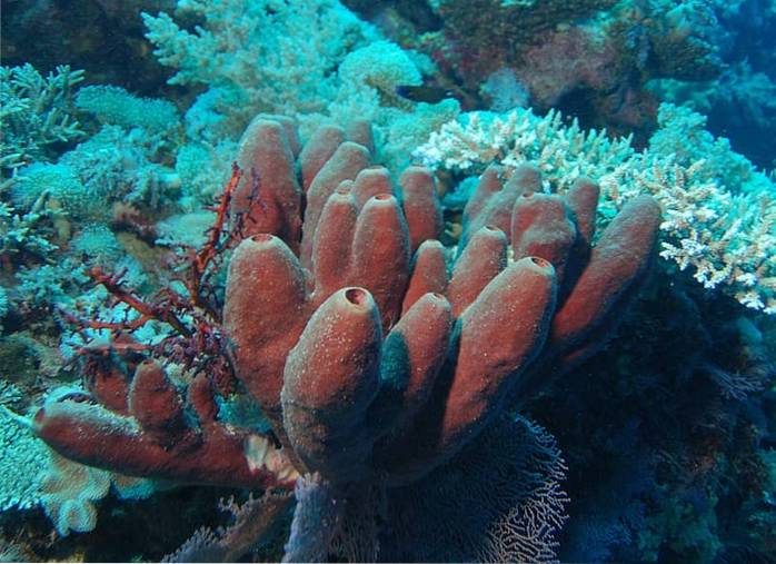
The choanocytes They are flagellate ovoid cells characteristic and exclusive of the Phylum Porífera, which use them to move water through a complex, also unique, of channels. These cells form a pseudoepithelium that lines the internal surfaces of the sponges that is known as coanoderm..
The coanoderm can be simple and continuous or acquire folds or subdivisions. In general, this pseudoepithelium consists of a single cell layer like the pinacoderm that lines the exterior.

Depending on the group of sponges, it can be folded or divided in some cases when the volume of the mesohilo of the sponge increases.
Article index
- 1 Features
- 1.1 Location of choanocytes
- 1.2 Asconoids
- 1.3 Siconoids
- 1.4 Leuconoids
- 2 Functions
- 2.1 Food
- 2.2 Playback
- 2.3 Excretion and gas exchange
- 3 References
Characteristics
In general they cover the atrium of the sponges and form chambers in the sponges of the group of syconoids and leuconoids..
The base of these cells rests on the mesohyl, which constitutes the connective tissue of sponges and its free end carries a contractile and transparent collar that surrounds a long flagellum at its base..
The contractile collar is made up of a series of microvilli, one next to the other that are connected to each other by thin microfibrils forming a mucous reticulum, forming a kind of highly efficient filtering device. The number of microvilli can be variable, however, it is between 20 to 55.
The flagellum has pulsating movements that attract water towards the microfibril collar and forces it to exit through the upper region of the collar that is open, allowing the entry of O2 and nutrients and the expulsion of waste.
Very small suspended particles are trapped in this network non-selectively. Those that are large slide through a secreted mucus towards the base of the collar where they are engulfed. Due to the role of choanocytes in phagocytosis and pinocytosis, these cells are highly vacuolated..
Location of choanocytes
The arrangement of the coanoderm determines the three body designs established within the porifers. These arrangements are directly related to the degree of complexity of the sponge. The flagellar movement of the choanocytes is not synchronized in any case, however, if they maintain the directionality of their movements.
These cells are responsible for generating currents within the sponges that pass through them completely through flagellar movement and the uptake of small food particles diluted in water or not, using phagocytosis and pinocytosis processes..
Asconoids
In asconoid sponges, which have the most simplistic design, the choanocytes are found in a large chamber called the spongiocele or atrium. This design has clear limitations since the choanocytes can only absorb food particles that are immediately close to the atrium..
As a consequence of this, the spongiocele must be small and therefore the asconoid sponges are tubular and small..
Siconoids
Although similar to asconoid sponges, in this body design, the internal pseudoepithelium, coanoderm, has folded outward to form a set of channels that are densely populated by choanocytes, thus increasing the absorption surface..
The diameter of these canals is markedly smaller compared to the spongiocele of asconoid sponges. In this sense, the water that enters the channels, a product of the flagellar movement of the choanocytes, is available and within reach to trap the food particles.
Food absorption only occurs in these channels, since the syconoid spongiocele does not have flagellate cells as in the asconoids and instead has covering cells of the epithelial type instead of choanocytes..
Leuconoids
In this type of body organization, the surfaces covered by choanocytes are considerably larger..
In this case, the choanocytes are arranged in small chambers where they can more effectively filter the available water. The body of the sponge has a large number of these chambers, in some large species it exceeds 2 million chambers..
Features
The absence of specialized tissues and organs in the Phylum Porífera implies that fundamental processes must occur at the individual cellular level. In this way, choanocytes can participate in various processes for the maintenance of the individual..
Feeding
Choanocytes obviously have an important role in sponge nutrition, as they are responsible for capturing food particles, using flagellar movement, the microvilli collar and the processes of phagocytosis and pinocytosis..
However, this task is not exclusive to the choanocytes and is also performed by cells of the outer epithelium, pinacocytes which engulf by phagocytosis food particles from the surrounding water and the totipotential cells of the porifers in the mesohilo (archaeocytes).
Within the choanocyte only a partial digestion of food occurs, since the digestive vacuole is transferred to an archaeocyte or other mesohyl wandering amoeboid cell where digestion ends.
The mobility of these cells in the mesohilo ensures the transport of nutrients throughout the body of the sponge. More than 80% of the nutritional material ingested is through the process of pinocytosis.
Reproduction
In addition, as far as reproduction is concerned, sperm appear to come from or originate from choanocytes. Similarly, in several species, choanocytes can also transform into oocytes, which also arise from archeocytes..
The process of spermatogenesis occurs when all the choanocytes in a chamber become spermagonia or when the transformed choanocytes migrate to the mesohyl and aggregate. However, in some demosponges the gametes originate from archeocytes..
After fertilization in viviparous sponges, the zygote develops within the parent, feeding on it, and then a ciliated larva is released. In these sponges, one individual releases the sperm and carries it to the channel system of the other..
There the choanocytes engulf the sperm and store it in food-like vesicles, becoming transporter cells..
These choanocytes lose their microvilli collar and flagellum, moving through the mesohyle as an amoeboid cell to the oocytes. These choanocytes are known as transference.
Gas excretion and exchange
The choanocytes also have a great participation in the processes of excretion and gas exchange. Part of these processes occur by simple diffusion through the coanoderm.
References
- Bosch, T. C. (Ed.). (2008). Stem cells: from hydra to man. Springer Science & Business Media.
- Brusca, R. C., & Brusca, G. J. (2005). Invertebrates. McGraw-Hill.
- Curtis, H., & Schnek, A. (2008). Curtis. Biology. Panamerican Medical Ed..
- Hickman, C. P, Roberts, L. S., Keen, S. L., Larson, A., I'Anson, H. & Eisenhour, D. J. (2008). Integrated Principles of zoology. McGraw-Hill. 14th Edition.
- Lesser, M. P. (2012). Advances in sponge science: physiology, chemical and microbial diversity, biotechnology. Academic Press.
- Meglitsch, P. A. S., & Frederick, R. Invertebrate zoology / by Paul A. Meglitsch, Frederick R. Schram (No. 592 M4.).


Yet No Comments