Coccidioides immitis is a dimorphic pathogenic fungus that causes an infection in the upper respiratory tract called coccidioidomycosis. This disease can present asymptomatic benign or moderately severe symptomatic. Rarely becomes fatal disseminated mycosis.
The fungus thrives in alkaline soils at extreme temperatures. For this reason, its habitat is described as a warm (54 ° C) and semi-arid environment (deserts with xerophilous vegetation). It is very tolerant of a wide variety of salt concentrations, including those that contain boron.
C. immitis It is found in endemic areas in the southwestern United States and northern Mexico. Some endemic foci are also observed in Central America, Venezuela, Colombia, Paraguay and Argentina.
Coccidioides immitis it is spread by airborne dust and its spores (arthroconidia) are naturally distributed thanks to air storms, when moving the earth or in excavations. These movements cause epidemics.
The fungus is concentrated in the entrances of rodent burrows, but it has not been possible to verify that there is an animal reservoir. The disease can affect both humans and some animals.
Coccidioidomycosis disease has a variety of alternative names, including: inn disease, coccidioidal granuloma, Valley fever, desert rheumatism, Valley bump, and California disease..
Article index
From childhood to puberty there are no differences in the rate of acquisition of the disease according to sex. However, in adulthood, men are more susceptible than women, with the exception of pregnant women who present the same risk as men. Obviously resistance to infection in women is linked to hormonal factors.
Likewise, race also influences the disease, with whites being the least susceptible, Indians and mestizos with moderate risk, and blacks the most affected by the disease, especially in disseminated cases..
Despite Coccidioides immitis it is considered the most virulent fungus of all the etiological agents of human mycoses, only 1% of primary infections develop into severe disease, and spread is 10 times more likely in the black race.
Of course, the infection is conditioned to the exposure of the fungus and the amount of the inoculum, and the risk increases in farmers, builders, archaeologists, among other occupations..
In the vast majority of patients, the primary disease is followed by recovery (without treatment) and the development of a specific immunity capable of protecting against reinfection..
People who develop disseminated infection are generally those who have a deficiency in their genetic or transient immune system.
Kingdom: Fungi
Division: Ascomycota
Class: Eurotiomycete
Order: Onygenales
Family: Onygenaceae
Gender: Coccidioides
Species: immitis
What Coccidioides immitis it is a dimorphic fungus, it presents with two morphologies. One saprophytic and the other parasitic.
In its saprophytic (infective) form, it is found as a mycelium, which presents septate hyphae, made up of chains of arthrospores or arthroconidia of a rectangular, ellipsoidal, barrel-like shape, with thick walls of 2.5 x 3-4 µ in diameter.
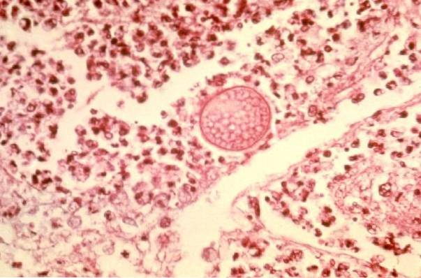
In its parasitic form it appears as a thick-walled spherule of 20 to 60 µ in diameter, filled with a large number of small endospores of 2-5 µ in diameter..
When these spherules break they release endospores (200 to 300) that can develop new spherules.
After 3 days of sowing a sample of infected tissue, you can see wet, glabrous or non-hairy colonies, later they are hairy, and later frankly cottony, grayish-white or yellowish..
Three types of reactions occur in infected tissues: purulent, granulomatous, and mixed..
The purulent reaction occurs initially around the inhaled conidia or at the time of rupture of the spherule and release of endospores.
The granulomatous reaction occurs around the developing spherule. The granuloma contains lymphocytes, plasma cells, monocytes, histiocytes, epithelioid cells, and giant cells.
These lesions then present fibrosis, caseification, and calcification. Later, in the lesions in which the microorganisms are growing and reproducing, the mixed reaction occurs.
The disease occurs after inhalation of dust containing arthroconidia. From there the disease can present itself in two ways.
The first asymptomatic or moderately severe, which will end with a complete remission of the infection and the development of permanent immunity.
The second is the rare form, where the disease progresses, becomes chronic or spreads, being fatal.
There are no symptoms, no residual scar, or lung injury, only the intradermal coccidioidin test is positive, indicating that there has been infection.
The intensity of the pathology will depend on the number of inhaled conidia. Few conidia will cause mild and brief illness, while a high inoculum can cause acute respiratory failure. On other occasions, it manifests itself with toxic erythema, arthralgia, episcleritis, etc..
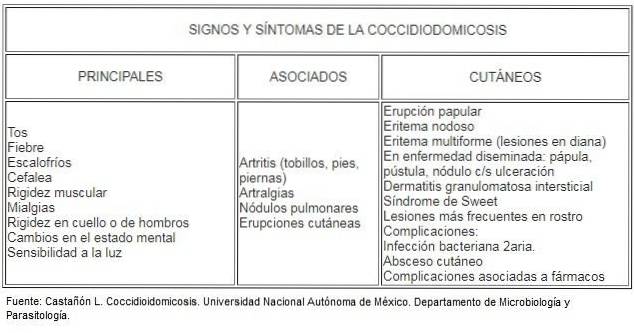
The incubation period is 10 to 16 days of incubation. After this time, patients may present to varying degrees the following signs and symptoms: fever, severe chest or pleuritic pain, respiratory distress, anorexia, initially non-productive cough and then productive with white sputum, and blood streaks.
It is very rare, caused by accidental inoculation of the fungus on the skin (prick with cactus spines). The lesion presents as a chancre, with regional adenitis, they subside without incident in a few weeks.
If the primary disease does not subside, after the sixth to eighth week, secondary or persistent manifestations will develop, which may present in two ways:
Secondary skin lesions are varied. They appear as: papules, nodules, warty, vegetating plaques, pustules, ulcers. They can be single or multiple.
They can also present as erythema nodosum, acute (“toxic”) rash, morbilliform erythema, interstitial granulomatous dermatitis, and Sweet's syndrome (febrile neutrophilic dermatosis)..
The fungus can also reach bones, joints, meninges and viscera. This type of coccidioidomycosis is fatal, causing the death of the individual in a few months to a year..
Other affectations resulting from a chronic residual coccidioidomycosis are cavitary disease and coccidioidoma.
Sputum, exudates, biopsies, CSF.
It is performed with the intention of finding spherules with typical endospores of coccidioidomycosis. These structures can be seen in tissue sections stained with hematoxylin and eosin, PAS, Gomori stain, Methanamine, silver nitrate, or calcium fluoride..
The samples are seeded on sabouraud or Mycosel agar, incubated at 25-30 ° C for 7 days. It is recommended to sow in tubes with slanted agar and not in a Petri dish..
For microscopic observation it is necessary to pass it previously through formaldehyde, to avoid accidental contamination. If subcultures are to be done, it must be under a security hood.
Complement fixation and precipitation reaction can be used. Diagnostic and prognostic value.
The intradermal coccidioidin reaction indicates whether the individual has been in contact with the fungus. Epidemiological value.
Although primary lung infection is usually self-limited in immunocompetent patients, it can be treated with itraconazole or fluconazole at doses of 400 mg per day for 3 to 6 months..
In immunosuppressed patients the same drugs are used but for 4 to 12 months.
In cases of chronic lung infection, fluconazole or itraconazole is used in doses of 400 mg per day for 12 to 18 months or more. Voriconazole has also given excellent results.
Amphotericin B is indicated for pregnant women.
Disseminated meningeal forms of coccidioidomycosis require lifelong treatment with fluconazole 400 mg per day.
In addition to antifungal therapy, surgical debridement of abscesses is indicated in some cases.


Yet No Comments