The Barr's corpuscle It is a mass of condensed heterochromatin that is observed within the female somatic cells of mammals and other animals. Generally easy to see during the mitotic interface stage.
Many scientists attribute this high concentration of heterochromatin to the inactivation of one of the two X chromosomes. This region stains intensely during cytological analyzes due to the large amount of heterochromatin it contains..
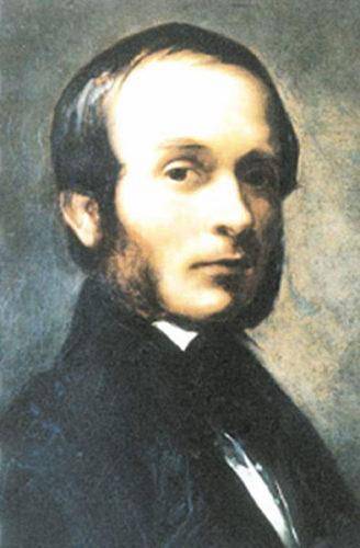
Barr's corpuscles were discovered by Murray Barr and Bertram in 1949. Both scientists observed that this small mass or body was present in the nuclei of nerve cells of domestic cats, while it was not evident in nerve cells of cats..
But it wasn't until 1966 that Mary Lyon proposed that these tiny corpuscles appeared as a result of the random inactivation of one of the two female sex chromosomes..
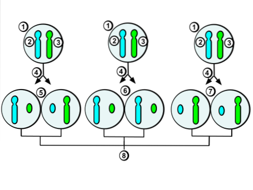
Many of the fertility problems diagnosed in women are due to the fact that their cells are in a "mosaic" form. This means that some of your cells do not inactivate one of your X chromosomes, but others do..
Thus, some cells have 45 somatic chromosomes and one active X sex chromosome, while others have 45 somatic chromosomes and two active XX chromosomes, which can have implications from many physiological and behavioral points of view..
Article index
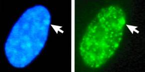
The Barr corpuscle is also called Barr's body or sexual heterochromatin. This is an element that has a circular, flat-convex shape when observed in the light microscope and has a length of approximately one micron..
Barr corpuscles, since they are composed of heterochromatin DNA, stain more intensely than echromatin DNA, which is "expanded" and dispersed within the cell nucleus.
Generally, hematoxylin and eosin are used for staining of this structure, which are compounds that stain cell nuclei blue, deep purple or black..
Barr's corpuscle is made up of facultative heterochromatin, that is, this DNA is expressed at certain times and not at others. When the DNA of the "active" or euchromatic X chromosome is defective, the DNA of the Barr corpuscle can become euchromatic to compensate for these failures..
In an average somatic cell, Barr's corpuscle is located on the inner face of the nucleus and, in Barr's first reports of the corpuscle, this structure is called a "nuclear satellite".
Digging deeper into his research, Barr found that these bodies were found in the cells of all female tissues, except for the cells of the liver and pancreas tissue..
In all mammals that develop through the placenta, there is an RNA in charge of initiating the silencing and packaging of the X chromosome that is not expressed, that is, of the formation of Barr's body. This RNA is called "X-specific inactive transcription RNA".
The "X-specific inactive transcription RNA" is only expressed to spread along the X chromosome selected by the cell to be silenced. The journey ends up stimulating cell silencing thanks to the participation of some of the histones present in the chromatin of said chromosome.
In order for the X-specific inactive transcription RNA to cover the entire chromosome length, cells must express between 300 and 1000 copies of it, so it has been found that there is a constant expression of the X-specific inactive transcription RNA to maintain to the second X chromosome in the form of a Barr body.
Scientists formerly proposed that "X-specific inactive transcription RNA" stimulated the formation of an internal repressive nucleus in Barr's corpuscle and that it contained a high content of repetitive DNA regions..
However, detailed observations with scanning electron microscopy have described Barr's corpuscle as a "suppressed" X chromosome that possesses highly packed chromatin, with loosely packed chromatin channels running from the periphery to the interior of the corpuscle..
All genes that control the chromosome silencing mechanism are conserved for all species, from yeast to humans. The complete locus that harbors these genes is called the "X-inactivation center.".
Murray Barr's discovery represented a breakthrough in making precise and detailed analyzes of the chromosomal sex of individuals. For example, for intersex disorders, Barr's body location and distinction soon became a widely used diagnostic tool..
This type of analysis is frequently carried out in forensic samples, since the chromatin of the X chromosome in its inactive form is present exclusively in female cells (remember that male cells also have an X chromosome, but this is active).
By extracting cells from human embryos, sex can be estimated early in development.
In addition, by identifying the sex, it is possible to diagnose diseases or abnormalities that are the product of the presence of more or fewer sex chromosomes than normal for cells in humans..
Individuals who possess two or more X chromosomes have a Barr body less than the number of X chromosomes within the cell nucleus. Thus, cells from abnormal females with a single X chromosome do not have any Barr corpuscles..
This anomaly is known as Turner syndrome; whereas cells from male individuals that have two XX chromosomes, one Y chromosome and a Barr body are diagnosed with Klinefelter syndrome.
There are also women who can have three X chromosomes and, therefore, have two Barr corpuscles inside the nuclei of their cells. However, cells that contain the abnormality for the sex chromosomes and cells that are completely normal can be found in the same person..
In general, individuals with these characteristics are sterile, have a “childish” appearance, which prevents them from having a full development, and are seen by some sectors of society as a kind of “phenomena”.
This is the condition referred to by "mosaic cells." People who do not have a total abnormality in their cells tend to have a lower degree of any of the syndromes.
During cytological analyzes, a tissue sample is quantified how many cells the abnormality has for its sex chromosomes; if the abnormality is in few cells, the individual may develop like a normal person.

Yet No Comments