A chromatid it is one of the two copies that is produced by the replication of a eukaryotic chromosome, visible only during cell division events such as mitosis and meiosis.
During mitotic division, sister chromatids are the result of DNA replication of the same chromosome and differ from homologous chromosomes in that they derive from two different individuals, a mother and a father, so, although they are recombine, they are not identical to each other.
Thus, chromatids are part of all eukaryotic chromosomes and fulfill essential functions in the faithful transfer of genetic information from a cell to its offspring, since the genetic content of two sister chromatids of a cell in mitosis, for example, is identical..
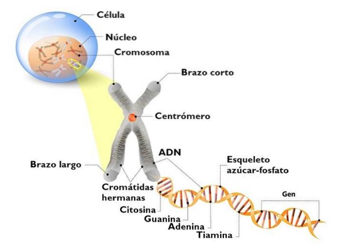
In general lines, each chromatid is composed of DNA wound on nuclei formed by octamers of histone proteins, which actively participate in the regulation of the expression of the genes contained in said DNA molecule..
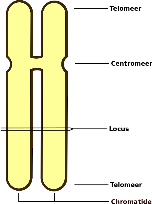
As they can only be seen during division, the chromatids are usually seen in pairs, closely attached to each other along their entire length and especially in the region of the centromere..
Article index
Chromatids are the nuclear structures that carry the genetic and epigenetic information of all eukaryotic cells. These are necessary for the correct distribution of hereditary material during cell division, either by mitosis or by meiosis..
Since the term is used especially to refer to the duplicated genetic material of a chromosome, a chromatid is therefore essential for the formation of genetically identical cells during mitosis and for the formation of gametes during meiosis of reproductive organisms. sexual.
The genetic material that is contained in the chromatids and that passes from a cell to its offspring through cell division contains all the information necessary to give the cells their own characteristics and, therefore, the organism that they form..
The proper segregation of sister chromatids is essential for the functioning of a living being, because if they are not transmitted faithfully from one cell to another or if they are not separated during division, genetic disorders that are detrimental to the development of the cell can be triggered. organism.
This is particularly true for diploid organisms such as humans, for example, but not entirely for other polyploid organisms such as plants, since they have “spare” sets of their chromosomes, that is, they have them in more of two copies.
Women, to name an example, have two copies of the sex chromosome X, so any replication error in one of them could be "corrected" or "amended" with the information present in the other, otherwise men, since they have a single copy of the Y chromosome and a single copy of the X chromosome, which are not homologous.
A chromatid is made up of a highly organized and compact double-band DNA molecule. The compaction of this molecule occurs thanks to its association with a set of histone proteins that form a structure called nucleosome, around which the DNA is wound.
The coiling of DNA around nucleosomes is possible because histones have abundant positively charged amino acids, which manage to interact electrostatically with the negative charges characteristic of nucleic acid..
The nucleosomes, in turn, roll on themselves, compacting even more and forming a filamentous structure known as the 30 nm fiber, which is the one observed during mitosis.
In a region of this card there is a DNA protein complex called the centromere, which houses the kinetochore, which is where the mitotic spindle binds during cell division..
At the end of the mitotic prophase it can be verified that each chromosome is composed of two filaments joined together throughout its entire structure and especially in a more compact region known as the centromere; these filaments are sister chromatids, the product of a previous replication.
The close union between sister chromatids throughout their structure is achieved thanks to a protein complex called cohesin, which functions as a "bridge" between the two. This cohesion is established as the DNA replicates, prior to the segregation of the chromatids towards the daughter cells..
When sister chromatids are separated during metaphase-anaphase, each chromatid that is secreted into one of the daughter cells is considered a chromosome, which replicates and forms a sister chromatid again before the next mitosis..
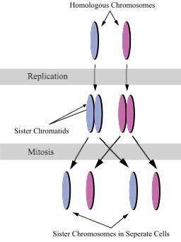
Most of the eukaryotic cells of organisms that have sexual reproduction have in their nucleus a set of chromosomes from one parent and another set from the other, that is, some chromosomes from the mother and others from the father, which are known as homologous chromosomes, as they are genetically equivalent, but not identical.
Each homologous chromosome is a highly ordered strand of DNA and proteins (chromatid) that, before the cell begins a division process, is loosely arranged in the nucleus.
Before a sex cell enters the meiotic phase, each homologous chromosome is duplicated, being composed of two identical sister chromatids joined throughout its structure and in the centromeric region, as happens during mitosis.
During the prophase of the first meiotic division, the homologous chromosomes (from the father and the mother), each already composed of two sister chromatids, approach each other along their entire length, through a process called synapse, by which a complex called a tetrad is formed, made up of each homologous chromosome and its sister chromatid.
The synapse allows genetic exchange or recombination between homologous chromosomes, which will later separate during anaphase I of meiosis and distribute into separate cells.
The sister chromatids of each homologous chromosome are secreted as a single unit during the first meiotic division, since they are displaced towards the same cell, but are separated from each other during meiosis II, where cells with a haploid number of chromosomes are produced..
Yet No Comments