The chronotropism It is the ability of heart cells to contract less or more frequently. It is considered one of the basic functional properties of the heart along with inotropism, dromotropism and bathmotropism.
Also know as rhythmicity, refers to the heart's ability to beat regularly. This phenomenon occurs thanks to the repetitive and stable depolarization and repolarization of cardiac muscle cells. As with inotropism, it is a generic term that over time became exclusively linked to the heart.
The word chronotropism has its etymological origin in ancient Greek. Chronos (chrónos) means "time." Trope (tropes) means "turn" or "turn". The ending "ism" is a typical noun former in the Greek language. Crono was the personification of the ages in Greek mythology, hence its use to refer to time.
Like all properties of the heart, chronotropism can be altered and cause disease. There are in turn several drugs that can modify the rhythm of the heartbeat, which in certain occasions can be considered harmful but in others it can have beneficial effects.
Article index
For a long time there was a controversy regarding the physiological origin of cardiac chronotropism. Why? Because some researchers suggested that the initial depolarization or "start" of the heartbeat was generated in the nervous tissue of the heart and another group claimed that it was produced from the muscle cell itself.
Today the myogenic theory is accepted over the neurogenic one. This decision is not whimsical but based on verifiable scientific facts, such as those mentioned below:
- Transplanted hearts beat regularly even when they are not connected to any nerves.
- In intrauterine life, the embryo's heart begins to beat before the nerve network develops.
- Some drugs are capable of inhibiting most of the body's nerves at certain doses, without affecting the heartbeat..
Ultimately, the rhythmicity of the heart is spontaneous and is due to the existence of an excitatory conductive system. This system is composed of non-contractile and self-excitable cardiac muscle cells. The role of the nerve network is limited to regulating the heart rate but not to start the beat.
The sinus node or sinoatrial node is the well-known natural pacemaker. This structure, made up of cardiomyocytes or cardiac muscle cells, is the site where the electrical impulse that causes the heartbeat is produced. Represents one of the fundamental structures of the electrical conduction system of the heart.
The sinus node is located in the muscular or myocardial wall of the atrium or right atrium. It is in immediate relation to the area of arrival of the superior vena cava. Some authors describe it in the shape of a banana and others assign it three recognizable parts: head, body and tail..
Its main function is to initiate the action potentials that will pass through the entire heart and cause the contraction or beat. The action potential is the change in the electrical charge of the cell membrane, which causes ion exchange and depolarization. The return to normal voltage across the membrane is known as repolarization..
The evaluation of chronotropism is achieved through the measurement of the heart rate. One of the fundamental characteristics of heart rhythmicity is that it is always generated, while the person is healthy, in the sinus node. This occurs because even with other pacemaker cells, those of the node are faster and opaque the rest..
The sinus node functions cyclically at the rate of 60 - 100 times per minute. This range represents the normal heart rate of a healthy adult. That is why measuring the number of beats in one minute is the easiest way to assess chronotropism. However, there are other ways to do it..
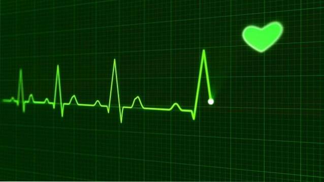
The electrocardiogram is a valuable classic. It allows to verify if the heart rate, even being within normal limits, has its origin in the sinus node.
The echocardiogram can also help in this task. Other more complex tests, such as cardiac electrophysiological studies, are useful for diagnosing rhythm disorders..
Chronotropism alterations are not always pathological. For example, high-performance athletes often have a slow heartbeat at rest, which is not considered abnormal..
Major physical effort or strong emotions can increase the heart rate, but this effect is physiological and does not require interventions.
- Sympathetic stimulation. The best example is the action of norepinephrine.
- Elevation of body or environmental temperature.
- Use of exogenous catecholamines or sympathomimetic drugs.
- Effects of thyroid hormones. Depending on the origin, it can be physiological (stress) or pathological (hyperthyroidism) events.
- Moderate hypoxia.
- Electrolyte disturbances. Hypocalcemia and hypokalemia can present with elevated heart rate in early stages.
- Vagal stimulation.
- Decreased body temperature.
- Use of cholinergic or parasympathomimetic drugs.
- Hypercapnia or elevated carbon dioxide. It can be generated by increased production or deficit elimination.
- Hydroelectrolytic alterations. Hyperkalemia, hypercalcemia, and hypernatremia.
- Diphtheria. In this case, it is the diphtheria toxin that causes, among other effects, a decrease in heart rate..
This group of drugs deserves a special mention. Digoxin, the main representative of digitalis, is one of the oldest known vasoactive drugs. It is obtained from foxglove plants or digitalis and has been used for centuries to treat some heart rate disorders.
Also known as cardiac glycosides, they are still widely used in the treatment of heart failure. The direct effects of these drugs are to increase the speed and force of the heartbeat. At high doses they can stimulate diuresis and increase peripheral resistance.
Digitalis poisoning is a serious and unfortunately common complication of the use of these drugs. The effect of intoxication is the opposite of its indication: it reduces the heart rate and can cause lethal arrhythmias. It also causes gastrointestinal complaints such as abdominal pain, nausea, vomiting, and diarrhea..

Yet No Comments