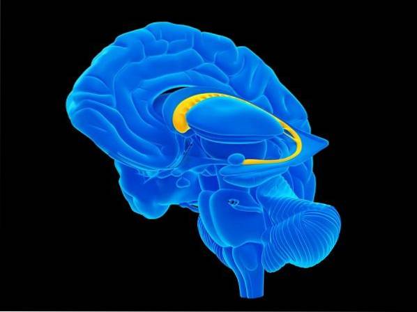
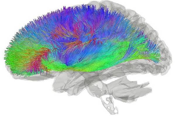
The striated body or striated nucleus is an important subcortical region that belongs to the forebrain. It is the main route of entry of information to the basal ganglia and is directly related to the cerebral cortex.
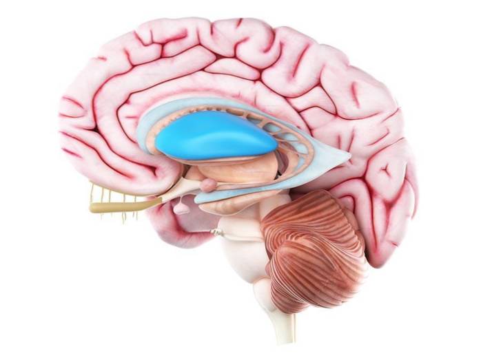
In humans, this brain structure is divided by a section of white matter known as the internal capsule. This section constitutes the two main structures of striated nuclei: the caudate nucleus and the lenticular nucleus..
Functionally, the striatum performs activities related to motor processes. In fact, it is part of the circuit known as the extrapyramidal system that is mainly responsible for regulating non-voluntary movements.
This article reviews the main characteristics of the striatum. Its anatomical properties and functions are discussed and the pathologies related to this structure of the brain are explained..
Article index
The striatum or rather, the striated nuclei because there is more than one, are a region of gray matter that is located inside the cerebral hemispheres. In this sense, they are subcortical structures that are located at the base of each hemisphere..
The two main nuclei that comprise the striatum are the caudate nucleus and the lenticular nucleus. The latter, in turn, is formed by two structures known as the putamen and the pale globe..
In this way, the striatum can be interpreted as a structure that encompasses different nuclei of the basal ganglia. These are:
- The caudate nucleus: structure linked to the processes of movement and learning.
- The putamen: linked structure in motor processes, operant conditioning and emotion regulation.
- The pale globe: structure that regulates the unconscious movements of the organism.
- The lenticular nucleus: region that is formed by the conjunction of the globe pallidus and the putamen.
On the other hand, in the ventral region, the striatum is made up of other structures. These are: the nucleus accumbens and the olfactory bulb.
Thus, this structure constitutes a large region of the brain that encompasses a large number of different structures and nuclei within it. It is a very important element of the brain as it establishes a constant connection with the cerebral cortex and the thalamic nuclei.
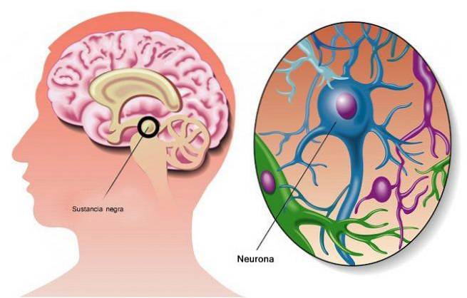
Likewise, the striatum is characterized by housing a large number of different neurons, such as medium spiny neurons, Deiter neurons, cholinergic inter-neurons or inter-neurons that express parvalbumin..
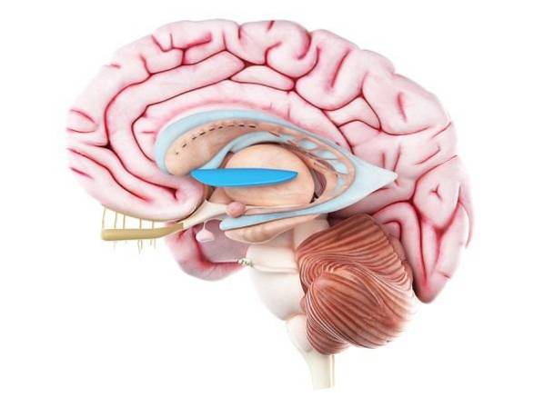
The striatum has a "C" shape when viewed from the side. The structure continues the course of the lateral ventricle and contains three main parts: the head, the body, and the tail..
Between the caudate and the putamen, two nuclei that are integrated into the interior of the striatum, a morphological continuity is observed. In fact, the anterior region of the caudate joins the head of the putamen.
The globe pallidus (another structure that is integrated into the corpus striatum) is found medial to the putamen. This nucleus has two regions: the lateral segment and the medial segment..
For its part, the caudate nucleus and the putamen also share a common embryological origin, as well as very similar connections. The set formed by these two structures within the striatum is called the neostriatum..
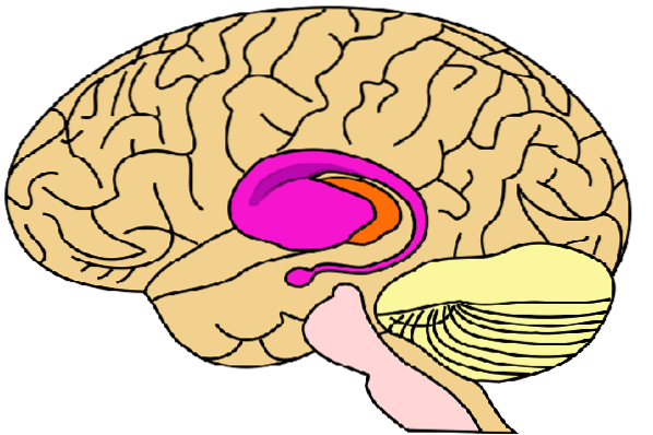
Finally, the putamen and globus pallidus form another "sub-group" within the striatum known as the lenticular nucleus..
All these nuclei, in turn, form part of the larger functional system of the basal ganglia system. This system is formed, beyond the striatum, by the sub-thalamic nucleus and the substantia nigra..
The striatum is characterized by being a very heterogeneous region in terms of the cell types that comprise it. Inside you can find many different types of neurons. These are:
They contain spines on the dendrites. These spinous cell extensions constitute practically the majority of the brain mass of the striatum (approximately 95%)..
They are characterized by having very long and little branched dendrites. They present a low prevalence within the stretched body, approximately 2%.
These cells are responsible for stopping electrical discharges in response to emotionally charged stimuli and elements related to gratification. They constitute 1% of the brain mass of the striatum.
They are responsible for emitting the substance parvalbumin. This substance, in turn, expresses receptors for catecholamines.
They are responsible for releasing a substance that is not very prevalent in the central nervous system known as calretinin..
These cells express somatostatin as well as dopamine receptors within the striatum..
The structures of the striatum communicate with different regions of the brain, encompassing both cortical and sub-cortical areas. These connections vary in each region of the striatum..
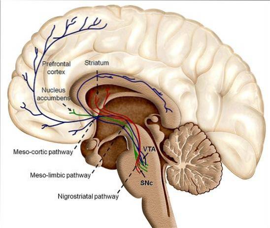
In this sense, the neostriatum (caudate and putamen) receive information from the cerebral cortex (mainly from the frontal lobe and the parietal lobe), from the substantia nigra that forms the negroestriate pathway, and from the intralaminar nuclei of the thalamus.
Likewise, these two structures of the striatum project their nerve fibers towards the pale nucleus and, on some occasions, to the substantia nigra..
The pale nucleus, on the other hand, receives nerve fibers from the neostriatum and the sub-thalamic nucleus. Its projections are directed towards the sub-thalamic nucleus and the thalamus.
The striatum is of high importance in motor circuits. Specifically, it is part of the extra-pyramidal system of the brain, which is responsible for regulating non-voluntary movements.
On the other hand, the putamen also seems to perform motor functions related to voluntary movements and the caudate is involved in cognitive activities.
Striatum disorders cause motor disturbances, such as involuntary movements, altered muscle tone, or tremors. In this sense, the two pathologies that have been associated with the functioning of this brain structure are: Parkinson's disease and Huntington's disease..


Yet No Comments