The embryonic and fetal development It is the process by which complete individuals are formed from cells from two parents: a father and a mother; corresponds to all the steps that follow the fertilization of an egg by a sperm, until birth.
The branch of medical sciences that is responsible for the analysis of these processes is known as "Embryology ”and its study began around 1651, when a scientist named Harvey realized that all individuals came from an“ egg ”.
However, the main advances in embryology did not take place until the arrival of the evolutionary concepts of Lamarck and Darwin in the 19th century, since before that time this science was supported by the "preformist" ideas of many scientists..
According to embryologists (the scientists in charge of the study of embryology), human development is divided into the prenatal and postnatal periods, which, as their names indicate, occur before and after birth, respectively..
Embryonic and fetal development correspond to the prenatal period, and it is the set of events in which the most drastic and important changes in development occur, since a fertilized cell called a zygote transforms into a highly complex multicellular organism.
It has been determined that the most obvious or visible changes occur between the third and eighth week of the embryonic period, while during fetal development there is growth and differentiation of the tissues and organs themselves.
The key processes that occur during embryonic and fetal development consist of multiple events of cell division, migration and programmed cell death, as well as cell ordering and complex information exchanges between cells.
Article index
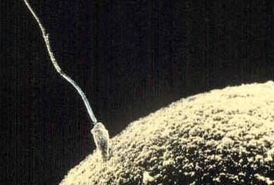
The embryonic development of any animal begins with the fertilization of an ovum by a sperm, which are the sex cells (gametes) of females and males, correspondingly.
In humans, this process occurs during the first 3 months (or the first 8 weeks) of gestation, after which the embryo is considered a fetus and, therefore, undergoes characteristic fetal development.
During the first week of gestation, the processes of fertilization and zygote formation take place; in this period the segmentation of this cell also occurs, producing the morula and blastula.
The fertilization process consists of a series of sequential events that are described from the first contact of the gametes to the fusion of their nuclei. These events can be listed as follows:
Note: once a sperm manages to "dissolve" the zona pellucida and reach the ovum, what embryologists have called a "reaction zone" is formed, which makes this cell impervious to other sperm.
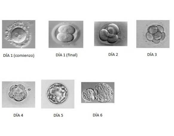
When the zygote is formed, that is, when fertilization has taken place and the chromosomal load has been restored, sequential mitotic divisions are triggered that achieve an increase in the number of cells (blastomeres).
Division involves a reduction in the size of the cells, but not an increase in volume, and occurs as the egg moves through the fallopian tubes toward the uterus. This process begins around 30 hours after fertilization..
When mitotic divisions complete around 12 or 32 cells (more or less on day 3 after fertilization), they “compact” thanks to adhesion events mediated by surface glycoproteins and form a “morula” (due to their morphological similarity with fruit).
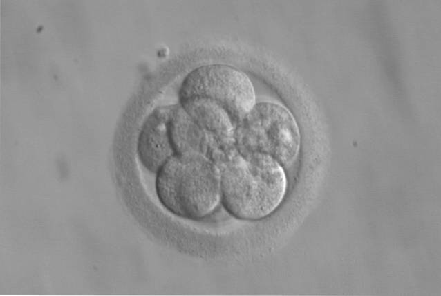
This morula is surrounded by a line of cells known as trophoblastic cells, which are the ones that will later form the placenta..
The consecutive divisions of the blastomeres of the morula generate a kind of cavity, the blastocele, which is why the resulting structure is known as a “blastula” or “blastocyst”. This structure is formed on day 4 after fertilization and when the morula reaches the uterus..
During the second week, in the blastocyst, two lines of cells begin to differentiate, each one coming from the cell lines originating from the two cells that are the product of the first division of the zygote..
One of the cell lines makes up the periphery of the blastocyst and is the one that will later give rise to the placenta, this layer is known as the trophectoderm.
The internal cell line, which surrounds the blastocelic cavity, corresponds to the organ-forming cells of the embryo that is in formation; in some books this layer of cells is known as embryoblastema or embryoblast.
It is between the 6th and 10th day when said blastocyst adheres to the endometrial epithelium, in the uterus, and it is there where the trophectoderm (also known as trophoblast) proliferates and differentiates into the cytotrophoblast (internal) and syncytiotrophoblast (external) layers..
All these processes are accompanied by abundant cell divisions and migrations, in addition to cell-cell adhesions or interactions that allow the formation of the mentioned layers..
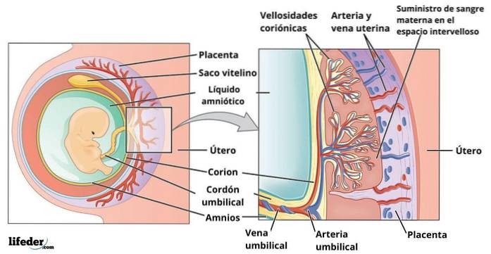
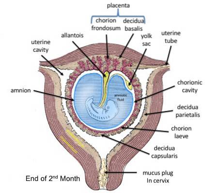
The second week of embryonic development is essential for the formation of the structures derived from the trophectoderm, that is, the “extra-embryonic” structures, which are: the amniotic cavity, the umbilical vesicle and the chorionic sac.
The third week is characterized by the differentiation of the three germ layers of the embryo during gastrulation; by the development of the notochord.
The blastomeres of the blastocyst continue to divide to form the gastrula, through the process known as gastrulation. At this stage of embryonic development, the fundamental embryonic “layers” begin to form..
Gastrulation also involves abundant cell migration, as well as their clumping and segregation. The gastrula is made up of an outer layer, the ectoblast or ectoderm, of a middle layer or mesoblast or mesoderm, and of an inner layer, the endoblast or endoderm..
At the end of the third week, the embryo has the appearance of a flattened, oval disk, in which the notochord between the ectoderm and the endoderm has already formed. The notochord is the primordial axis of the embryo, around which the axial skeleton is formed, that is, it is a "proto-vertebral column".
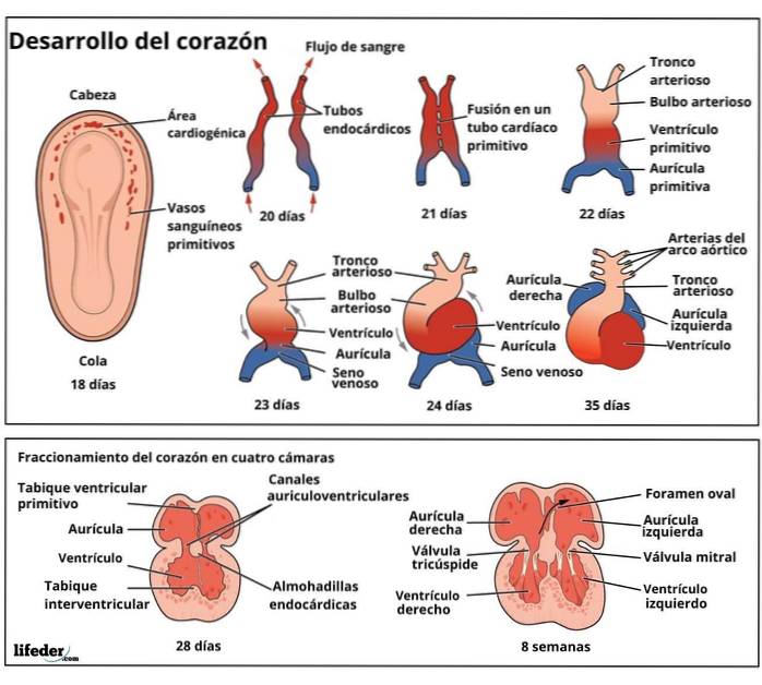
Likewise, in this stage, the neural plate, neural folds and neural tube are formed in the ectoderm, which correspond to the primordium of the central nervous system. The primordia of the cardiovascular system is also outlined during the third week.
The main internal and external structures are formed between the fourth and eighth weeks of embryonic development. During these weeks, the processes of growth, morphogenesis and differentiation of tissues and organs take place..
These processes are finely regulated and controlled, especially by the genetic expression patterns of the cells that are part of the germ layers in question, which depend, to some extent, on environmental characteristics..
The body shape of the embryo originates from the folding of the trilaminar, oval, and discoidal embryo that formed during the third week. This process occurs in the middle and horizontal plane of the same and after this the embryo grows relatively quickly.
In the process of folding, the primordium of the brain, pharynx, esophagus and lower respiratory system are formed. Part of the endodermal layer is used for the formation of the hindgut, the descending colon, and the rectum..
Although the progression from the embryo to the fetus occurs gradually, the distinction is necessary to affirm that, in the fetus, the structures of a growing human being are recognized, since the main organs and body systems have already been formed.
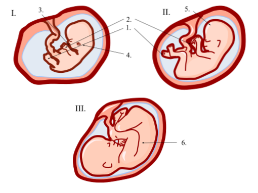
The fetal period begins in the ninth week of gestation. Between the ninth and twelfth weeks the growth of the fetus accelerates, but a disproportionate relationship with the body and head remains.

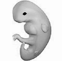
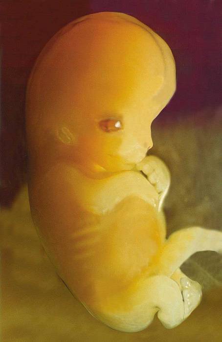
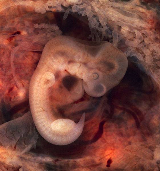
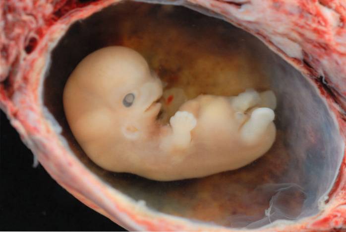
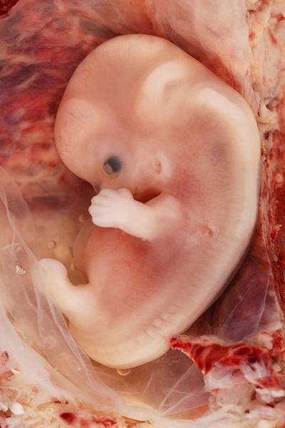
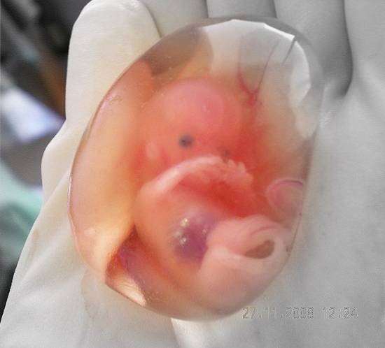
During the ninth week the distinctive features are: a very wide face, wide-set eyes, fused eyelids, and "drooping" ears. The legs are short and the thighs are relatively small. In the following image you can see an embryo at 9 weeks of gestation:
Until the end of the ninth week the external genitalia are indistinguishable between boys and girls. In a nine-week fetus, the liver is the main site for red blood cell formation (erythropoiesis) and it is during this time that urine begins to form..
By the time the fetus reaches the twelfth week, the primary ossification centers appear in the skull skeleton and long bones. In addition, in this period, the upper limbs reach their relative definitive lengths, but the lower limbs still have to develop..
Between these weeks, growth accelerates even more and becomes more evident. At the end of 16 weeks the body acquires a size more proportional to that of the head and the lower limbs have reached their corresponding length.
Between these 3 weeks, the true ossification of the skeleton begins and the development of the bones can be observed on ultrasound. By week 14, slow eye movements can be seen and the scalp pattern is also determined..
From these weeks, sex can be determined, since in females the ovaries and primordial germ cells differentiate. In addition, the eyes are no longer located anterolaterally and are arranged in the anterior region of the face..
The ears also settle into their final positions on the sides of the head.

The growth rate slows a little after week 17, but during this time interval fetal movements begin to become evident..
Between weeks 17 and 20, the skin of fetuses is covered with a protective waxy substance called “waxy vernix”, and also with a thin layer of hairs (lanugo) that contributes to the adhesion of the vernix to the skin.
During this time, the eyebrows and hair become visible and the brown fat begins to be deposited, which participates in the production of heat..
The fetus, with wrinkled and pink skin, begins to gain weight. He has rapid eye movements and his lungs begin to produce pulmonary surfactant. Fingernails usually appear during week 24.
By the end of these three weeks, the fetus already has a sufficiently developed pulmonary system to carry out gas exchange.
The eyes are open, the hair has developed, and the toenails are also visible. In addition, the fetus increases the synthesis of white fats, which results in a gain in body mass..
At the end of week 28, the bone marrow takes over the production of red blood cells, which previously occurred in the spleen and before there, in the liver.
At week 30, the development of the pupillary reflex or, what is the same, the change in the diameter of the pupil in response to light has been documented. By this time the percentage of body fat is greater than 7% and the fetus's extremities appear plump..
From this point it is considered that the gestation is in a period of termination. Fetuses born prematurely, from week 26, have a chance to survive with medical assistance, but from week 35 onwards they are less at risk.
Characteristics such as the relationship between the circumferences of the head and the abdomen or the length of the feet are used during this period to determine the age of the fetus..
At week 38, a full-term pregnancy is already considered. During this time the percentage of body fat is approximately 16% and the chest and pectorals protrude slightly in both boys and girls..


Yet No Comments