The ectoderm it is one of the three germ layers that appear in early embryonic development. The other two are the mesoderm and endoderm, which lie below this.
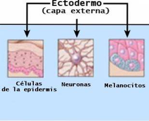
The ectoderm or outer layer gives rise, mainly, to the nervous system, epidermis and associated structures such as hairs and nails. It is present in the development of practically all living beings.
This germ sheet is the first to develop, appearing in the blastula stage. The blastula is an early phase in which the embryo has about 70 to 100 cells that can develop into any type of tissue. It appears between 4 to 6 days after fertilization, and is sometimes used as a synonym for ectoderm.
Before being trilaminar, the embryo has two layers: the hypoblast and the epiblast. The ectoderm arises from the epiblast. During the next phase, called gastrulation, this layer gives rise to the endoderm and the mesoderm through the invagination of cells.
Each of these layers will give rise to different types of cells that will make up various parts of the body, as well as the umbilical cord, placenta and amniotic fluid..
The next period of embryonic development is known as neurulation. This stage begins with a thickening of the ectoderm in the dorsal midline. This is due to a very important structure located immediately below the ectoderm, called the notochord..
This structure is responsible for sending inductive signals to the ectoderm so that it accumulates cells and is invagine. In addition, it will induce a part of your cells to differentiate into nerve precursor cells, which will make up the nervous system..
This thickening of the ectoderm is known as the "neural plate." As neurulation progresses, the neural plate thickens while a crack appears in its middle to invaginate. The neural plate is the precursor to the neural crest and neural tube, which are discussed later..
The term ectoderm comes from the Greek "έξω" or "ektos", which means "outside" and "δέρμα" or "dermis", which means "skin".
Article index
In vertebrate organisms, three important parts can be distinguished in the ectoderm:
This area is the one that gives rise to the epithelial tissues such as the glands of the skin, the mouth, the nasal cavities, the hair, the nails, part of the eyes, etc. In animals, it produces feathers, horns and hooves.
As mentioned before, the ectoderm undergoes thickening during the neurulation phase. It will accumulate cells that are arranged in two chains, on both sides of the midline of the neural plate.
At 20 days of gestation, the neural plate begins to fold in its midline, giving rise to the neural groove, which deepens each time. Thus, the structure invaginates to form the neural tube.
The area of the neural plate that lies above the notochord is called the floor plate. While, the area furthest from the notochord is known as the neural crest. This is located at the most dorsal limit of the neural tube, and is a group of cells that appears in the region where the edges of the folded neural plate meet..
Neural crest cell subsets migrate following pathways in which they receive additional inductive signals that will influence their differentiation. Therefore, these cells will become a great variety of structures.
There are four different migration pathways for the differentiation of neural crest cells. Each pathway determines what specific cell structures they will transform into. Thus, they will lead to:
- The neurons and glial cells of the sensory ganglia, which are fundamental components of the peripheral nervous system.
- The neurons and glia of the autonomic ganglia, which include the ganglia of the sympathetic and parasympathetic nervous system.
- Neurosecretory cells of the adrenal glands, which are included in the dorsal part of the kidneys.
- Cells that are going to transform into non-neural tissues, such as melanocytes. The latter have the objective of producing melanin in the skin. There are also groups of cells that will make up the cartilage of the face and teeth..
The neural tube closes like a zipper. It begins in the cervical region, and from there it continues in a cranial and caudal direction. Until the fusion is complete, the cranial and caudal ends of the neural tube remain open, communicating with the amniotic cavity.
When the most cranial end is closed, dilations called brain vesicles appear. These are the ones that will give rise to the brain, specifically its first divisions: the rhombencephalon, the midbrain and the forebrain..
Whereas, the most caudal and narrow part of the neural tube will become the spinal cord. In the case in which the cranial neuropore does not close, the encephalic vesicles will not develop.
This causes a very serious condition called anencephaly, which prevents the brain and skull bones from forming. If the neural tube of the ectoderm closes poorly, the individual may present with spina bifida..
On the other hand, the cells of the neural tube will also constitute the retina of the eyes and the neurohypophysis. The latter is the posterior lobe of the pituitary gland..
The last two parts are called the neuroectoderm.
The ectoderm derives in the following structures:
- Nervous system (brain, spinal cord, and peripheral nerves).
- Epidermis.
- Sweat and mammary glands.
- Toothpaste.
- Lining of the mouth, nostrils, and anus.
- Hair and nails.
- The lenses of the eyes.
- Inner ear parts.
Ectodermal dysplasia is a rare but serious disease that arises from a mutation or combination of mutations in several genes..
Thus, genes do not give the correct signals for the ectoderm to develop as it should. In this disease it is observed that several tissues derived from the ectoderm do not form properly. For example, teeth, skin, hair, sweat glands, nails, etc..
Actually, there are more than 170 subtypes of ectodermal dysplasia. The most common type is hypohidrotic ectodermal dysplasia, which is characterized by hypohidrosis or the inability to sweat (due to malformation of the sweat glands).
It is also usually accompanied by facial malformations, such as missing teeth, wrinkled skin around the eyes, a deformed nose, eczema on the skin, and scant and fine hair..
It has been observed that this subtype is hereditary, following a recessive pattern linked to the X chromosome. It occurs more in males, since they only have one X chromosome..


Yet No Comments