
The term ectrodactyly is used to call a rare inherited syndrome, in which there is malformation of the fingers and toes. It is characterized by the absence of the distal phalanges or complete fingers.
It can occur in one or more fingers of the hand and even affect part of the carpus and wrist. In severe cases, and when the condition affects the foot, the fibula or all four limbs may be absent..
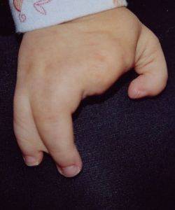
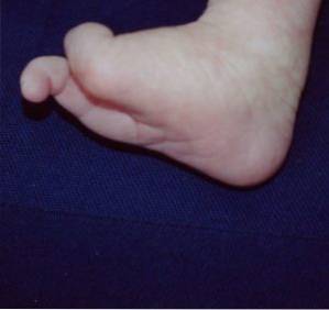
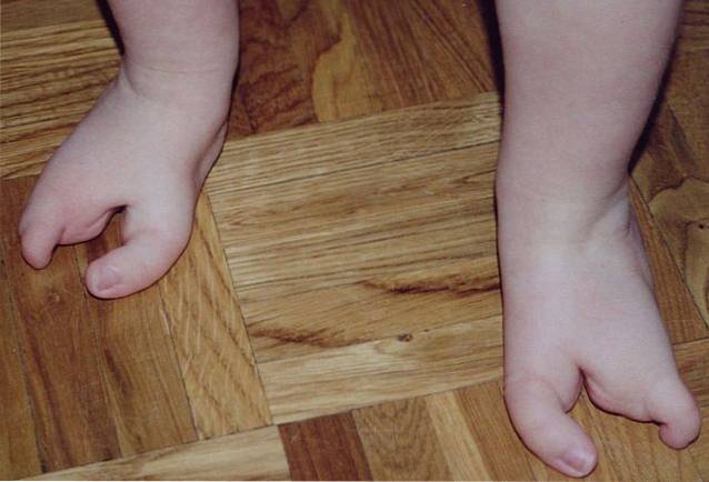
In the most common types of ectrodactyly, the third finger of the hand is missing, creating a deep cleft in that space. The remaining fingers are attached by soft tissues. This is known as syndactyly, and it is what ends up giving the look of lobster claw.
The syndrome includes other deformities such as cleft lip and palate, tear duct obstruction, and genito-urinary defects..
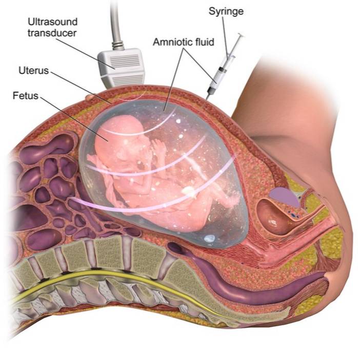
Prenatal diagnosis is made through the evaluation of the fetus's limbs by ultrasound. If the condition is confirmed, consultation with specialists in genetic counseling is recommended for the chromosome study of the parents..
Although there is no treatment for this disease, there are conservative and surgical measures that are used to improve the quality of life of the patient..
Article index
Also know as Karsch-Neugebauer syndrome, ectrodactyly is a rare condition characterized by deformity of the hands and feet. 1 in 90,000 people suffers from this disease.
Although sporadic cases have been described, these are very rare. Ectrodactyly is described as a hereditary disease that originates from the mutation of one of the genes on chromosome 7, which is the one most frequently involved in this disease.
There are two types of ectrodactyly, type I is the most common; in this there is malformation of the hands and / or feet without any other bodily alteration.
In contrast, type II is more serious and rare. In these patients, a cleft palate is observed, in addition to the characteristic malformations. There may also be visual and genito-urinary system problems.
The patient with ectrodactyly has a very characteristic deformity in the hands and feet. Depending on the severity of the disease, and according to the individual's genetic mutation, different malformations are observed.
In type I ectrodactyly, abnormalities are observed in the fingers and toes, although they can occur in only one limb, their course is unpredictable.
The typical characteristic in these patients is the absence of the middle finger with the union of the remaining fingers on each side..
At the site of the absent finger, a deep cleft can be seen, giving the appearance of lobster claw. This is the same in the hands and in the feet.
In severe cases there may be absence of more fingers and even bones of the carpus, wrist, fibula or the four limbs.
Type II ectrodactyly occurs with other malformations, in addition to those seen in this syndrome.
It is commonly associated with a cleft palate and lip, partial or complete absence of teeth, tear duct abnormalities, photophobia, and decreased visual acuity. Genito-urinary malformations such as an underdeveloped kidney, among others, can also be observed..
In pregnancy control evaluations, the specialist doctor is able to observe a facial or limb malformation with ultrasound, from 8to gestation week.
When the characteristic malformation of ectrodactyly is evident, the disease should be suspected even if it is not present in the family.
The definitive prenatal diagnosis is made from the genetic study of the amniotic fluid, which is obtained through a procedure called amniocentesis, which generally poses no danger to the mother or fetus.
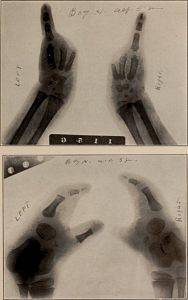
After birth, the malformations caused by the genetic mutation are evident, so the suspected diagnosis is made from the physical examination.
Conventional radiographs of the hands and feet are used to observe the patient's skeleton, which is sometimes underdeveloped or with fetal remnants of primitive bone..
This disease does not have a treatment aimed at its cure. However, measures are taken so that the patient improves their quality of life and can relate to their environment in a normal way..
Thus, there are conservative techniques and surgical techniques that help the individual with ectrodactyly to carry out their daily tasks without problems..
Conservative treatment includes the use of prostheses and special insoles that improve gait and stability..
In the case of hand deformity, physical therapy and rehabilitation are the most helpful elements for the patient..
For its part, surgical treatment is used to optimize finger movements, improve gait and avoid foot deformity that limits the use of footwear..
In all cases, the fingers are separated with syndactyly and the cleft is closed, which helps to maintain the shape of the foot and hand..
Doctors specializing in genetics have a special advisory and advice service for patients who are carriers or have diseases that can be transmitted to their children..
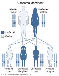
In the case of ectrodactyly, it is inherited in a dominant way. That is, the children of gene carriers have a 50% chance of suffering from the disease.
Genetic counseling is responsible for explaining this form of transmission, in addition to the probabilities that children have the disease.
Ectrodactyly is a condition that causes a great psychological impact for both the patient and their families, even generating rejection of the patient.
Genetic counseling seeks to prepare parents for this situation, in case the diagnosis of the disease has been confirmed in an unborn child.
Genetic counseling is a fundamental service in the case of inherited diseases.


Yet No Comments