
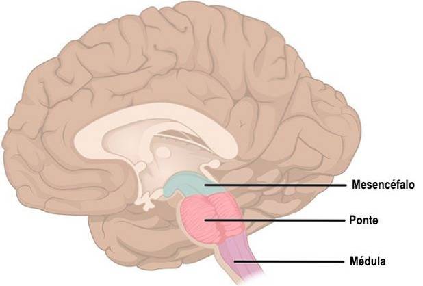
The bridging or brainstem bridge is a region of the brain stem that in humans is located in the lower midbrain, in front of the cerebellum and directly above the medulla oblongata. It was the Italian anatomist Varolio who named this structure as such, which is why it is also known as “Varolio Bridge”. This scientist perceived this region as such due to its prominence and the similarity it had to a bridge that connected the two hemispheres of the cerebellum.
Contents
The brainstem bridge is the most prominent area of the brainstem. The shape of the brainstem bridge resembles the middle of a ring that connects the left and right cerebellum from its sides, as well as the cerebellum with the midbrain. Despite this, the bridge is not a direct connection between these two hemispheres. In this structure, two areas are distinguished, one posterior and one anterior..
The posterior area of the bridge gives rise directly to the cerebellum and connects to the medial cerebellar peduncles, large bundles of fibers that connect the bridge to the cerebellum, being one of the main connection routes of the brain with this structure. In addition, from this area the bridge forms the base of the fourth ventricle.
The anterior or rostral part contains transverse fibers and is crossed by the basilar artery, one of the sources of the highest proportion of oxygenated blood in the brain.
Different very important cranial nerves arise from the bridge that connect with other areas of the brain. These are the trigeminal nerve, which has sensory and motor functions, the abducens nerve, which is involved in eye movements, the facial nerve, which manages to control the muscles of the face creating facial expressions, and the vestibulocochlear nerve, which is related to the auditory information. Thus, the nuclei that make up the brainstem bridge are associated with these nerves, being: the abductor somatic motor nucleus, the special trigeminal motor nucleus, the special facial motor nucleus and the superior salivatory nucleus..
Internally, the bridge is made up of the ventral area, in which the pontine nucleus is located, which is responsible for coordinating movement. In the tegmentum, the oldest part of the bridge, which is part of the reticular formation, there are a set of nuclei responsible for the arousal system and care.
The bridge contains groups of neurons essential to the neurotransmitter systems of the nervous system. For example, the locus coeruleus that is found in the dorsal part of the bridge and that is involved in stress processes, gives rise to the largest collection of noradrenergic neurons in the nervous system that project to the rest of it. The nuclei of the Rafe, also located in the bridge are groups of neurons that contain a high level of serotonin.
As we have seen, the brainstem bridge is a structure composed of several nuclei and tracts that have very diverse functions and are involved in numerous actions, from facial expressions to aspects related to sleep, especially with the REM phase of it. The brainstem bridge becomes an area of connection between different areas of the brain that cross messages from the cortex or the cerebellum, connecting the upper and lower areas of the brain.
Thus, the bridge functions as a connecting center that intervenes in the general functioning of the nervous system and whose functions address different capacities, both motor and regulatory, ranging from balance, through breathing, to emotions..
When injuries to the brainstem bridge occur, the consequences affect multiple functional capacities that are very basic for human survival. The injuries that can occur in this area are normally caused by vascular problems, as well as tumors or trauma, although different diseases can also affect its function, such as Parkinson's disease or Alzheimer's.
Millard-Gübler syndrome is a disease caused by an injury to the lower bridge that can cause facial palsy ipsilateral to the injury, as well as contralateral brachiocrural palsy. This injury is usually caused by vascular causes, although it can also be due to a tumor or trauma. Thus, a paralysis occurs on the side opposite to the injury that affects the face or body of the affected person..
Inferior Foville syndrome is also due to injury to this region of the brain, usually caused by problems such as tumors or heart attacks. In this case, the affected fibers are the VI and VII of the cranial nerves and ocular paralysis occurs, as well as contralateral pyramidal hemiplegia and ipsilateral facial paralysis. This can also lead to deafness or dizziness, as well as loss of taste since, as we see, the implications of the bridge are so wide that its injuries can affect very different capacities..


Yet No Comments