The encephalocele, Cephalocele, craniocele or bifid skull is a disease that arises from a defect in the neural tube (NTD). It occurs very early, when the embryo is developing inside the uterus; And what happens is that the cells that make up the skull do not unite as they should, so that they leave part of the brain outside.
It consists of a series of congenital malformations that arise during the embryonic stage in which the skull does not close and part of the brain tissues protrudes from it. It can have different locations in the skull of the affected person, which will influence the diagnosis, treatment and progression of the disease.
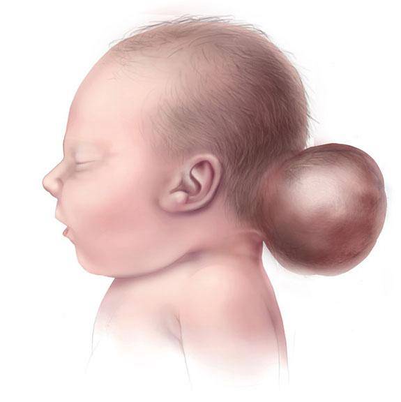
It is an uncommon defect, manifested by the naked eye as a sac-like protrusion outside the skull, which is normally covered by a thin membranous layer or skin..
It then consists of a defect in the bones of the skull that causes part of the meninges (membranes that cover the brain), brain tissue, ventricles, cerebrospinal fluid or bone to protrude from it..
It can occur in any area of the head; but the most common is in the middle posterior part (in the middle of the occipital area of the brain). When the encephalocele is in this place, neurological problems usually appear.
Approximately half of those affected by encephalocele will present a significant cognitive deficit, mainly motor learning, although it depends on the brain structures that are affected.
This condition is often diagnosed before or immediately after birth as it is highly visible, although there are extremely rare cases in which the bump is very small and can be missed..
Article index
The exact cause that causes encephalocele to appear is still unknown, although it is thought that it is surely due to the joint participation of several factors; such as the mother's diet, the exposure of the fetus to toxic or infectious agents, or even genetic predisposition.
This condition is more common in people who already have a family history of diseases linked to neural tube defects (such as spina bifida), so it is suspected that genes may be involved.
However, someone who has a genetic predisposition to certain disorders may carry a gene or genes associated with the disease; but it is not necessarily going to develop it. It seems that environmental factors also have to contribute. In fact, most cases occur sporadically..
All these factors would cause a failure in the closure of the neural tube during the development of the fetus. What allows the brain and spinal cord to form is the neural tube.
It is a narrow canal that must be folded in the third or fourth week of pregnancy for the nervous system to build properly. The bad closure of the neural tube can occur anywhere in it and therefore there are types of encephalocele with different locations.
There are specific factors that have been associated with this disease, such as the mother's lack of folic acid. In fact, it appears that the rate of encephalocele decreases as fertile women do not have dietary folic acid deficiencies..
Encephalocele can go hand in hand with more than 30 different syndromes, such as Fraser syndrome, Roberts syndrome, Meckel syndrome, amniotic band syndrome, or Walker-Warburg syndrome, Dandy-Walker syndrome, Chiari malformation ; among others.
As for future pregnancies, if an isolated encephalocele occurs; there is no risk of future pregnancies with the same condition. However, if it is part of a syndrome with several associated anomalies, it may be repeated in future children..
Encephalocele can be classified into different types depending on the tissues involved:
- Meningocele: only part of the meninges protrude.
- Encephalomeningocele: contains meninges and brain tissue.
- Hydroencephalomeningocele: it is more severe, as tissues of the brain protrude including ventricles in addition to part of the meninges.
As we mentioned, they are also classified by their location. The most common places where encephalocele develops are:
- Occipital: at the back of the skull.
- The upper middle zone.
- Frontobasal: between the forehead and the nose, which can be divided into nasofrontal, nasoethmoidal or nasorbital.
- Sphenoid or by the base of the skull (involving the sphenoid bone)
Encephalocele is a very rare condition, occurring in approximately 1 in 5,000 live births worldwide. It seems to be commonly associated with stillbirth before 20 weeks' gestation, while only 20% are born alive.
In fact, according to the Metropolitan Atlanta Congenital Defects Program (Siffel et al., 2003), the majority of deaths in children with encephalocele occurred during the first day of life and the estimated probability of survival to 20 years of age was of 67.3%.
It seems that other malformations and / or chromosomal abnormalities may appear in at least 60% of patients with encephalocele..
Occipital encephaloceles occur most frequently in Europe and North America, while frontobasal encephaloceles are more common in Africa, Southeast Asia, Russia, and Malaysia..
According to the “Centers for Disease Control and Prevention” (2014), women belonging to the female sex are more likely to develop encephalocele in the back of the skull, while in men it is more likely in the frontal part.
The symptoms of an encephalocele can vary from one individual to another depending on many different factors, including the size, location, and the amount and type of brain tissue that protrudes from the skull..
Encephaloceles are usually accompanied by:
- Craniofacial malformations or brain abnormalities.
- Microcephaly, or reduced size of the head. That is, its circumference is smaller than expected for the baby's age and sex..
- Hydrocephalus, which means a buildup of excess cerebrospinal fluid, putting pressure on the brain.
- Spastic quadriplegia, that is, progressive weakness of the muscles due to increased tone that can lead to paralysis or total loss of strength in the arms and legs.
- Ataxia (incoordination and voluntary motor instability).
- Developmental delay, including growth and mental retardation that prevent you from learning normally and reaching developmental milestones. However, some affected children may have normal intelligence..
- Vision problems.
- Seizures.
However, it is essential to point out that not all affected individuals will present the aforementioned symptoms..
Today, most cases are diagnosed before birth. Mainly through a routine prenatal ultrasound, which reflects sound waves and projects the image of the fetus.
Encephalocele can appear as a cyst. However, as we said, some cases can go unnoticed; especially if they are on the forehead or near the nose.
It should be borne in mind that the ultrasound appearance of an encephalocele can vary during the first trimester of pregnancy..
Once an encephalocele is diagnosed, a careful search should be made for possible associated abnormalities. For this, other additional tests can be used, such as prenatal MRI that offers more details.
Here are the tests that can be used for the diagnosis and evaluation of this disease:
- Ultrasound: Your resolution of the fine details of the brain and / or CNS is sometimes limited by the physical makeup of the mother, the surrounding amniotic fluid, and the position of the fetus. However, if it is 3D ultrasound; Liao et al. (2012) point out that this technique can help early detection of encephalocele in the fetal stage, providing a vivid visual representation, which contributes significantly to the diagnosis.
- Bone scan
- Magnetic resonance imaging: may lead to better results than fetal ultrasound, as the central nervous system of the fetus can be viewed in great detail and non-invasively. However, it requires anesthesia of the mother and the embryo. It can be useful in babies with this problem, also perform it after birth.
- Computed tomography: although it has sometimes been used for the early diagnosis of encephalocele and its associated problems, radiation in fetuses is not recommended; mainly in the first 2 trimesters of pregnancy. Best used after birth, as they provide a good representation of bone defects in the skull. However, it is not as effective as magnetic resonance imaging (MRI) in representing soft tissues..
- Nuclear imaging, such as nuclear ventriculography or radionuclide cisternography. The latter is useful for observing the circulation of cerebrospinal fluid, and they are carried out by injecting radioactive substances as markers and then observing them circulate in the body through an imaging technique such as SPECT or single-photon emission computed tomography..
- Angiography: it is mainly used to evaluate intracranial and extracranial vascular aspects, and is usually used before performing a surgical intervention. Its use is recommended if there is a concern about the probable venous displacement of the pons. However, its use to evaluate an encephalocele is infrequent, since magnetic resonance imaging can also allow observation of the venous anatomy..
- Amniocentesis can also be performed to detect potential chromosomal abnormalities or implications..
On the other hand, a genetic consultation is recommended in any family that has a baby affected by encephalocele.
Surgery will usually be used to place the protruding tissue inside the skull and close the opening, as well as correct the craniofacial malformations. The protrusions can even be removed without causing major disabilities. Possible hydrocephalus is also corrected through surgical treatment.
However, according to Children's Hospital of Wisconsin, It should be mentioned that this surgery is not usually performed immediately after birth, but rather waits for a while; which can range from days to months, so that the baby adjusts to life outside the uterus before opting for the operation.
Thus, the vast majority of surgical interventions are carried out between birth and 4 months of age. However, the urgency of the surgery will depend on several factors depending on the size, location and complications that it entails. For example, it should be operated urgently if there is:
- Lack of skin covering the sac.
- Exsanguination.
- Airway obstruction.
- Vision problems.
If it is not urgent, the baby will be thoroughly examined for other abnormalities before surgery is performed..
Regarding the surgical procedure, first, the neurosurgeon will remove a part of the skull (craniotomy) to access the brain. He will then cut the dura, the membrane that covers the brain, and correctly position the brain, meninges, and cerebrospinal fluid in place, removing the excess sac. Later, the dura will be closed, sealing the extracted part of the skull or adding an artificial piece to replace it..
On the other hand, hydrocephalus can be treated with an implantation of a tube in the skull that drains excess fluid..
Another additional treatment, depends on the symptoms of each individual and can be simply symptomatic or supportive. When the problem is very severe and / or is accompanied by other alterations; palliative care is commonly recommended.
That is, you will take care of yourself, feed yourself and give you oxygen to increase your maximum comfort level; but no attempt will be made to extend the life of the baby with life support machines.
Parental education is very important for the treatment, being part of local, regional and national associations and organizations can be of great use and relief..
In terms of prevention, studies have shown that adding folic acid (a form of vitamin B) to the diet of women who want to become pregnant in the future can lower the risk of neural tube defects in their children. . It is recommended for these women to ingest an amount of 400 micrograms of folic acid per day.
Other important factors to prevent encephalocele are taking health measures before and after pregnancy such as quitting smoking and eliminating alcohol consumption..
The prognosis of this disease depends on the type of tissue that is involved, where the sacs are located and the consequent malformations that occur.
For example, encephaloceles located in the frontal area tend not to contain brain tissue, and therefore have a better prognosis than those located in the back of the skull. That is, the absence of brain tissue within the pons is an indicator of better results, as well as the absence of associated malformations..
In the "Centers for Disease Control and Prevention" (2014) they investigate what may be the risk factors for encephalocele, finding for now that children with this disease have a lower survival rate and: multiple birth defects, low newborn weight born, preterm birth, and being black or African American.


Yet No Comments