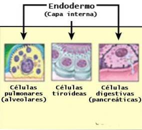
The endoderm it is one of the three germ layers that arise in early embryonic development, around the third week of gestation. The other two layers are known as the ectoderm or outer layer and the mesoderm or middle layer. Below these would be the endoderm or inner layer, which is the finest of all.
Before the formation of these layers, the embryo is composed of a single sheet of cells. Through the gastrulation process, the embryo invaginates (folds back on itself) to produce the three primitive cell layers. First the ectoderm appears, then the endoderm and finally the mesoderm.
Before gastrulation, the embryo is just a layer of cells that later divides into two: the hypoblast and the epiblast. On day 16 of gestation, a series of migratory cells flow through the primitive streak, displacing the cells of the hypoblast to transform into the definitive endoderm.
Later, a phenomenon called organogenesis occurs. Thanks to this, the embryonic layers begin to change to become the different organs and tissues of the body. Each layer will give rise to different structures.
In this case, the endoderm will originate the digestive and respiratory systems. It also forms the epithelial lining of many parts of the body..
However, it is important to know that what they form are rudimentary organs. That is, they do not have a specific shape or size and have yet to fully develop..
At first the endoderm is made up of flattened cells, which are endothelial cells that mainly form lining tissues. They are wider than they are tall. Later they develop into columnar cells, which means they are taller than they are wide.
One of the oldest layers of embryonic differentiation in living things is the endoderm. For this reason, the most important organs for the survival of the individual come from it..
Article index
The differentiation of the body of the embryo from the external fluid affects the endoderm, dividing it into two parts: the embryonic and extra-embryonic endoderm.
However, the two compartments communicate by a wide opening, a precursor of the umbilical cord.
It is the part of the endoderm that will form structures within the embryo. Gives rise to the primitive intestine.
This germ layer is responsible, together with the mesoderm, for originating the notochord. The notochord is a structure that has important functions. Once formed, it is located in the mesoderm, and is responsible for transmitting inductive signals for cells to migrate, accumulate and differentiate..
The transformation of the endoderm parallels the changes induced by the notochord. Thus, the notochord induces folds that will determine the cranial, caudal and lateral axes of the embryo. The endoderm also folds progressively into the body cavity under the influence of the notochord.
At first it begins with the so-called intestinal sulcus, which invaginates until it closes and forms a cylinder: the intestinal tube..
The other portion of endoderm is outside the embryo, and is called the yolk sac. The yolk sac consists of a membranous structure attached to the embryo that is responsible for nourishing, giving it oxygen and eliminating waste.
It only exists in the early stages of development, up to about the tenth week of gestation. In humans, this sac acts as the circulatory system.
On the other hand, different areas can be differentiated in the intestinal tube of the endoderm. It should be said that some of them belong to the embryonic endoderm and others to the extra-embryonic:
- The cranial or inner intestine, which is located within the fold of the embryo's head. It begins in the oropharyngeal membrane, and this region goes on to become the pharynx. Then, at the lower end of the pharynx, a structure appears that will originate the respiratory tract.
Below this area, the tube will quickly widen to later become the stomach..
- Middle intestine, located between the cranial and caudal intestines. This is extended to the yolk sac through the umbilical cord. This allows the embryo to receive nutrients from its mother's body..
- The caudal intestine, within the caudal fold. From it arises the allantois, an extra-embryonic membrane that appears by an invagination located next to the yolk sac.
It consists of a deposit that leaves the embryonic body through the allantoic pedicle (umbilical cord). The volume of fluid in the bag changes as gestation progresses, since it seems that this sac accumulates metabolic waste.
In humans, the allantois gives rise to the umbilical vessels and the villi of the placenta.
As mentioned, the endoderm derives organs and structures in the body through a process called organogenesis. Organogenesis occurs in a stage that lasts from the third to the eighth week of gestation approximately.
The endoderm contributes to the formation of the following structures:
- Glands of the gastrointestinal tract and associated gastrointestinal organs such as the liver, gallbladder, and pancreas.
- Surrounding epithelium or connective tissue: tonsils, pharynx, larynx, trachea, lungs, and gastrointestinal tract (minus the mouth, anus, and part of the pharynx and rectum, which come from the ectoderm).
It also forms the epithelium of the Eustachian tube and the tympanic cavity (in the ear), the thyroid and parathyroid glands, the thymus gland, the vagina, and the urethra..
- Respiratory tract: such as bronchi and pulmonary alveoli.
- Urinary bladder.
- Yolk sac.
- Allantois.
It has been shown that in humans the endoderm can differentiate into observable organs after 5 weeks of gestation.
The ectoderm changes by the induction of the notochord at first, and later by a series of growth factors that regulate its development and differentiation..
The whole process is mediated by complex genetic mechanisms. Therefore, if there are mutations in an associated gene, genetic syndromes may appear in which certain structures do not develop properly or present malformations. In addition to genetics, this process is also sensitive to harmful external influences.
Different investigations have identified these proteins as markers for the development of the endoderm in various species:
- FOXA2: it is expressed in the previous primitive line to build the endoderm, it is a protein encoded in humans by the FOXA2 gene.
- Sox17: plays an important role in the regulation of embryonic development, especially in the formation of the endoderm intestine and the primitive heart tube.
- CXCR4: or type 4 chemokine receptor, is a protein that in humans is encoded by the CXCR4 gene.
- Daf1 (complement deactivation accelerating factor).

Yet No Comments