The endonucleases they are enzymes that cut the phosphodiester bonds located inside the nucleotide chain. Endonuclease restriction sites are highly varied. Some of these enzymes cut DNA (deoxyribonucleic acid, our genetic material) almost anywhere, that is, they are nonspecific.
In contrast, there is another group of endonucleases that are very specific in the region or sequence that they are to cleave. This group of enzymes is known as restriction enzymes, and they are very useful in molecular biology. In this group we have the well-known enzymes Bam HI, Eco RI and Alu I.
Contrary to endonucleases, there are another type of catalytic proteins - exonucleases - that are responsible for breaking the phosphodiester bonds at the end of the chain.
Article index
Restriction endonucleases or restriction enzymes are catalytic proteins that are responsible for cleaving the phosphodiester bonds inside the DNA chain in very specific sequences.
These enzymes can be purchased from multiple biotechnology companies and their use is almost essential within current DNA manipulation techniques..
Restriction endonucleases are named using the first letters of the binomial scientific name of the organism they come from, followed by the strain (this is optional) and ending with the group of restriction enzymes to which they belong. For example, Bam HI and Eco RI are widely used endonucleases..
The region of DNA that the enzyme recognizes is called the restriction site and is unique to each endonuclease, although several enzymes may coincide at the restriction sites. This site generally consists of a short palindromic sequence about 4 to 6 base pairs in length, such as AGCT (for Alu I) and GAATTC for Eco RI..
Palindromic sequences are sequences that, although read in the 5 'to 3' or 3 'to 5' direction, are identical. For example, for the case of Eco RI, the palindromic sequence is: GAATTC and CTTAAG.
Fortunately for molecular biologists, bacteria have developed in the course of evolution a series of restriction endonucleases that internally fragment genetic material.
In nature, these enzymes have evolved - presumably - as a bacterial protection system against the invasion of foreign DNA molecules, such as those from phages.
In order to discriminate between native and foreign genetic material, these restriction endonucleases can recognize specific nucleotide sequences. Thus, DNA that does not have this sequence can be undisturbed inside the bacterium..
In contrast, when the endonuclease recognizes the restriction site, it binds to the DNA and cuts it..
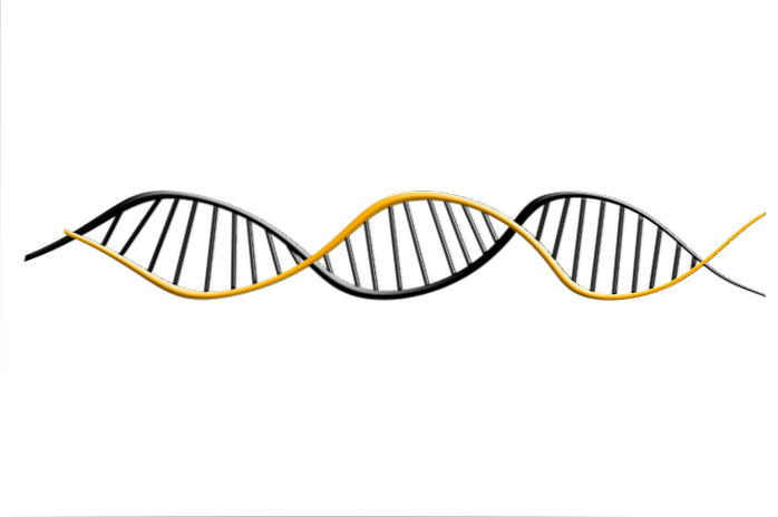
Biologists are interested in studying the genetic material of living things. However, DNA is made up of several million base pairs in length. These molecules are extremely long and must be analyzed in small fragments..
To meet this goal, restriction endonucleases are integrated into various molecular biology protocols. For example, an individual gene can be captured and replicated for future analysis. This process is called "cloning" a gene..
Restriction fragment length polymorphisms refer to the pattern of specific nucleotide sequences in DNA that restriction endonucleases are able to recognize and cut.
Thanks to the specificity of the enzymes, each organism is characterized by a specific pattern of cutting in the DNA, originating fragment of variable lengths.
Historically, restriction endonucleases have been classified into three types of enzymes, designated by Roman numerals. Lately, a fourth type of endonuclease has been described.
The most important characteristic of type I endonucleases is that they are proteins made up of several subunits. Each of these functions as a single protein complex and usually have two subunits called R, two M and one S.
The S portion is responsible for the recognition of the restriction site in DNA. The R subunit, for its part, is essential for cleavage and M is responsible for catalyzing the methylation reaction..
There are four subcategories of type I enzymes, known by the letters A, B, C, and D, that are in common use. This classification is based on genetic complementation.
Type I enzymes were the first restriction endonucleases to be discovered and purified. However, the most useful in molecular biology are type II, which will be described in the next section..
Type II restriction endonucleases recognize specific DNA sequences and cleavage at a constant position near a sequence that produces 5 'phosphates and 3' hydroxyls. They generally require magnesium ions (Mgtwo+), but there are some that have much more specific requirements.
Structurally, they can appear as monomers, dimers or even tetramers. Recombinant technology uses type II endonucleases and for this reason more than 3,500 enzymes have been characterized.
These enzyme systems are made up of two genes, called mod Y beef, that code for the subunits that recognize DNA and for modifications or restrictions. Both sub-cities are necessary for restriction, a process totally dependent on the hydrolysis of ATP.
In order to cleave the DNA molecule, the enzyme must interact with two copies of the non-palindromic recognition sequence and the sites must be in a reverse orientation on the substrate. Cleavage is preceded by a DNA translocation.
An additional group has been identified lately. The system is composed of two or more genes that code for proteins that cleave only modified DNA sequences, either methylated, hydroxymethylated or hydromethylated glucosyl.
For example, the enzyme EckKMcrBC recognizes two dinucleotides of the general form RmC; a purine followed by a methylated cytosine, which can be separated by several base pairs - from 40 to almost 3000. Cleavage takes place about 30 base pairs behind the site that the enzyme recognizes.
Endonucleases of this type are also known as endonucleases "homing”. These enzymes recognize and cut the target DNA sequence at unique sites in the genome from 14 to 40 bp.
These enzymes are often encoded in introns and their function is believed to be to promote horizontal transfer of the cut sequences. After cutting, a break repair occurs in the DNA double helix based on the complementary sequence.
Endonuclease I of E. coli it acts as a defense system against phages and parasites. It is located mainly between the cytoplasmic membrane and the cell wall. Produces double-stranded breaks in the foreign DNA with which it interacts in the periplasmic space.
CRISPR-Cas endonucleases are enzymes that act on the defense mechanism of many types of bacteria. These identify and cut specific DNA sequences from invading organisms, which are generally viruses.
Recently, researchers from the Massachusetts Institute of Technology (MIT) discovered the CRISPR-Cas12bm genomic editing system with high precision for the modification of human cells..


Yet No Comments