The endospores They are forms of survival of certain bacteria, made up of dehydrated sleeping cells covered by protective layers, which show extreme resistance to physical and chemical stress. They are capable of lasting indefinitely in the absence of nutrients. They form inside bacteria.
Endospores are the most resistant living structures known. They can survive high temperatures, ultraviolet light, gamma radiation, desiccation, osmosis, chemical agents, and enzymatic hydrolysis.
When environmental conditions determine it, endospores germinate giving rise to active bacteria that feed and multiply.
Endospores are a type of spore. There are fungi, protozoa, algae, and plants that produce their own types. Endospores lack reproductive function: each bacterial cell produces only one. In other organisms, on the contrary, they can have reproductive function.
Article index
In the middle of the 17th century, the Dutch cloth merchant and precursor of microbiology Antonie van Leeuwenhoek, using ingenious microscopes designed and made by himself, was the first to observe living microorganisms, including protozoa, algae, yeasts, fungi and bacteria..
In 1859, the French Academy of Sciences sponsored a competition in which the French chemist Louis Pasteur participated. The objective was to shed light through an experiment on "spontaneous generation", an ancient hypothesis that proposed that life can arise from "vital forces" or "transmissible substances" present in non-living or decomposing matter..
Pasteur showed that, as in the case of wine, air and solid particles are the source of the microbes that grow in culture broths previously sterilized with heat. Shortly thereafter, in 1877, the English physicist John Tyndall corroborated Pasteur's observations, giving the final blow to the hypothesis of spontaneous generation..
Tyndall also provided evidence for extremely heat-resistant forms of bacteria. Independently, between 1872 and 1885, the German botanist Ferdinand Cohn, considered the founder of modern microbiology, described bacterial endospores in detail..
Most organisms live in environments that vary in time and space. A common strategy to survive environmental conditions temporarily unsuitable for growth and reproduction is to enter a state of reversible dormancy, during which individuals take refuge in protective structures and minimize their energy expenditure..
The transition between active and latent states is metabolically costly. This investment is greater when individuals must build their own protective structures, be they composed of exogenous materials, or biosynthesized within. In addition, individuals must be able to respond to environmental stimuli that cause the transition..
Dormancy generates a reservoir of dormant individuals that can be activated when favorable conditions reappear. These reservoirs allow the conservation of populations and their genetic diversity. When it comes to pathogenic endospore-producing bacteria, latency facilitates their transmission and makes their control difficult..
Bacterial endospores can remain viable for many years. It has been argued that endospores preserved in ancient substrates, such as permafrost, aquatic sediments, underground salt deposits, or amber can remain viable for thousands and even millions of years..
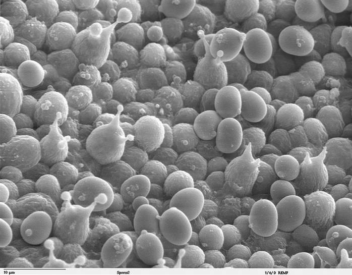
Visualizing the position and other characteristics of the endospores is very useful for the identification of species of bacteria..
Endospores can be seen using a light microscope. In bacteria subjected to Gram or methylene blue staining, these are distinguished as colorless regions within the vegetative bacterial cell. This is because the endospore walls are resistant to penetration by ordinary staining reagents..
A specific staining method for endospores, known as the Schaeffer-Fulton differential stain, has been developed which makes them clearly visible. This method allows to visualize both those that are inside the bacterial vegetative cell and those that are outside it.
The Schaeffer-Fulton method is based on the ability of malachite green to stain the wall of endospores. After applying this substance, safranin is used to color the vegetative cells.
The result is a differential staining of endospores and vegetative cells. The former acquire a green color and the latter a pinkish color..
Within the vegetative cell, or sporangium, the endospores can be located terminal, subterminal, or centrally. This bacterial form has four layers: medulla, germ wall, cortex, and cover. In some species there is a fifth outer membranous layer called exosporium, composed of lipoprotein that contains carbohydrates.
The medulla or center is the protoplast of the endospore. It contains the chromosome, ribosomes, and a glycolytic energy-generating system. May not have cytochromes, even in aerobic species.
The energy for germination is stored in 3-phosphoglycerate (there is no ATP). It has a high concentration of dipicolinic acid (5-15% of the dry weight of the endospore).
The germ wall of the spore surrounds the medullary membrane. Contains typical peptidoglycan, which during gemination becomes the cell wall of the vegetative cell.
The cortex is the thickest layer of the endospore. Surrounds the germ wall. Contains atypical peptidoglycan, with less cross-links than typical, which makes it very sensitive to autolysis by lysozymes, necessary for germination.
The coat is composed of a keratin-like protein that contains numerous intramolecular disulfide bonds. Surrounds the cortex. Its impermeability confers resistance to chemical attacks.
Dipicolinic acid appears to play a role in latency maintenance, DNA stabilization, and heat resistance. The presence of small soluble proteins in this acid saturates the DNA and protects it from heat, desiccation, ultraviolet light and chemical agents..
The synthesis of the atypical peptidoglycan begins when an asymmetric septum is formed that divides the vegetative cell. In this way, the peptidoglycan divides the stem cell in which the prespore will develop into two compartments. Peptidoglycan protects it from osmotic imbalances.
The cortex osmotically removes water from the protoplast, making it more resistant to heat and radiation damage..
Endospores contain DNA repair enzymes, which act during marrow activation and subsequent germination.
The process of forming an endospore from a vegetative bacterial cell is called sporulation or sporogenesis..
Endospores occur more frequently when certain critical nutrients are in short supply. There may also be production of endospores, which represent life insurance against extinction, when nutrients are abundant and other environmental conditions are favorable..
Sporulation consists of five phases:
1) Formation of the septum (medullary membrane, germ wall of the spore). A portion of the cytoplasm (future medulla) and a replicated chromosome are isolated.
2) The germ wall of the spore develops.
3) The cortex is synthesized.
4) The cover is formed.
5) The vegetative cell degrades and dies, thus releasing the endospore.
The process by which an endospore transforms into a vegetative cell is called germination. This is triggered by the enzymatic breakdown of the endospore covering, which allows the hydration of the marrow and the restart of metabolic activity..
Germination consists of three phases:
1) Activation. Occurs when abrasion, a chemical agent, or heat damage the cover.
2) Germination (or initiation). It starts if the environmental conditions are favorable. Peptidoglycan is degraded, dipicolinic acid is released, and the cell is hydrated.
3) Outbreak. The cortex is degraded and biosynthesis and cell division are restarted.
The endospores of pathogenic bacteria are a serious health problem due to their resistance to heating, freezing, dehydration and radiation, which does kill vegetative cells.
For example, some endospores can survive for several hours in boiling water (100 ° C). In contrast, vegetative cells do not resist temperatures above 70 ° C.
Certain endospore-producing bacteria of the genera Clostridium Y Bacillus excrete potent protein toxins that cause botulism, tetanus, and anthrax.
Depending on the case, treatments include gastric lavage, wound cleansing, antibiotics, or antitoxin therapy. Preventive measures include hygiene, sterilization and vaccination.
It is caused by contamination with spores of Clostridium botulinum. Its most obvious symptom is muscle paralysis, which can be followed by death. Its incidence is low.
There are three types of botulism. The infantile is caused by the ingestion of honey or other additives, contaminated by air, that have been added to the milk. For its part, food is produced by ingestion of contaminated food (such as canned food), raw or poorly cooked. Finally, the injury is produced by contact with the earth, which is the natural habitat of C. botulinum.
It is caused by Clostridium tetani. Its symptoms include muscle contractions that are very painful (in Greek, the word "tetanus" means to contract) and so strong that they can cause broken bones. It is often fatal. Its incidence is low.
Infective spores of C. tetani they typically enter the body through a wound, in which they germinate. During growth, which requires that the wound is not well oxygenated, the vegetative cells produce the tetanus toxin.
The bacteria and its endospores are common in the environment, including soil. They have been found in the feces of humans and animals.
It is caused by Bacillus anthracis. Its symptoms vary greatly depending on the environment and site of infection. It is a serious and often fatal disease. Its incidence is moderately high, leading to epidemics in animals and humans. In the 18th century, anthrax decimated Europe's sheep.
Herbivorous mammals are its natural host. Humans become infected by contact (usually occupational) with animals, or by handling or ingesting animal products.
There are three types of anthrax:
1) Cutaneous. The entrance is produced by injuries. Blackish, necrotic ulcers form on the skin.
2) By inhalation. Entrance during breathing. Produces inflammation and internal bleeding and leads to coma.
3) Gastrointestinal. Entry by ingestion. Produces oropharyngeal ulcers, severe abdominal bleeding and diarrhea.
In approximately 95% of cases, human anthrax is cutaneous. In less than 1% it is gastrointestinal type.
Endospores can be destroyed by sterilization in autoclaves, combining pressures of 15 psi and temperatures of 115-125 ° C for 7-70 minutes. They can also be eliminated by alternating changes in temperature and pressure, such that there is germination of spores followed by death of the resulting vegetative bacteria..
Peracetic acid is one of the most effective chemical agents for destroying endospores. Iodine, tinctured (dissolved in alcohol) or iodophor (combined with an organic molecule) is also often lethal to endospores..
The destruction of endospores in surgical instruments is effectively achieved by introducing them into a container into which a plasma (excited gas rich in free radicals) is induced, for which certain chemical agents are subjected to negative pressure and an electromagnetic field..
The destruction of endospores in large objects, such as mattresses, is achieved by exposing them for several hours to ethylene oxide combined with a non-flammable gas..
Food processing industries use chlorine dioxide in aqueous solution to fumigate areas potentially contaminated with endospores of anthrax..
Sodium nitrite added to meat products, and the antibiotic nisin added to cheese, prevent the growth of endospore-producing bacteria.
Bacillus anthracis it is easy to grow. For this reason, during the two world wars it was included as a biological weapon in the arsenals of Germany, Great Britain, the United States, Japan and the Soviet Union..
In 1937 the Japanese army used anthrax as a biological weapon against Chinese civilians in Manchuria. In 1979, in Sverdlovsk, Russia, at least 64 people died from accidentally inhaling spores from a strain of B. anthracis of military origin. In Japan and the United States, anthrax has been used for terrorist purposes.
In contrast, attempts are currently being made to use endospore coatings as a vehicle for therapeutic drugs and for antigens created for preventive immunization purposes..


Yet No Comments