The Entamoeba hartmanni is a species of amoeba belonging to the genus Entamoeba, considered non-pathogenic, does not have an invasive stage, nor does it consume red blood cells in the way that E. histolytica or E. dispar are distinguished.
This species has been the subject of various debates since 1912, when the scientist Prowazek detected small cysts smaller than 10mc under a microscope. He classified them as a new species of Entamoeba and named it Hartmanni. On the other hand, Wenyon and Col determined that it was a small race belonging to E. histolytica, although at present it is not disputed that it is a new species.
In this sense, the determination of the methods for the diagnosis and characterization of the morpho-genetic aspects, as well as the transmission mechanisms, contagion symptoms, standardized or special treatments, are of vital importance for an adequate understanding of this organism belonging to the order Entamoebida.
Article index
-Entamoeba hartmanni, like the other amoebae, biologically belongs to the eukaryotic domain and is classified within the protist kingdom.
-This amoeba has a vacuolated cytoplasm, a unique and differentiated nucleus that shows a central endosome in trophozoites.
-Peripheral chromatin shows a homogeneous distribution throughout the body.
-Another interesting aspect is that they do not engulf erythrocytes. The oligonucleotide sequence in Entamoeba hartmanni is;
GTGAAGAGAAAGGATATCCAAAGT (AF149907)
Fundamentally, the morphological characteristics of this amoeba are found in its stages, two of which are;
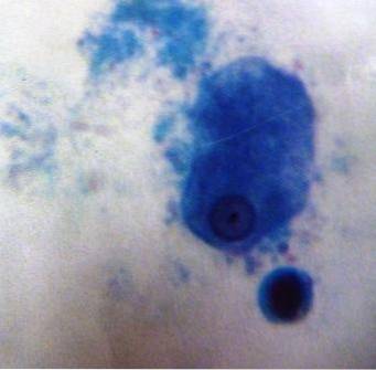
During this phase, the organism has a rounded or amoeboid shape and a size that ranges from 5 to 12 μm, with an average of 8 to 10 μm. Its movement, in general, does not turn out to be progressive and the only nucleus that it presents is not visible when observed in preparations without dyeing..
In properly stained samples it is possible to observe a karyosome of small proportions, compact and located in the central area. However, on various occasions it may be outside the center.
In the same way, it contains perinuclear chromatin, which takes the form of tiny and fine granules of uniform size and distribution, although a beaded shape can sometimes be present..
Also, the cytoplasm is thinly granular and can usually contain some bacteria, but never shows the presence of red blood cells. This is due to your inability to ingest them..
They have a generally spherical shape, with a diameter that varies from 5 to 10 μm, being regularly between 6 and 8 μm.
In this sense, the most mature cysts manifest 4 nuclei, not visible when the samples observed through microscopy are not properly stained..
By having Lugol's staining in proportions of 20.gm of I2 and 40.gm of KI correctly dissolved in 1.Lts of H2O, it is possible to observe them. Also, undeveloped cysts, with 1 or 2 nuclei, are more common in tests than mature cysts..
When seen in stained preparations, the nuclei have a small central karyosome and regularly distributed perinuclear chromatin with fine, uniform grains..
Also, in the same way that it happens with the other species of the “Entamoeba complex”, glycogen can be little differentiable and dispersed in mature cysts..
However, in immature cysts it is more concise and the chromatoidal bodies can acquire a cluster shape, as well as be elongated with slightly rounded ends..
Non-pathogenic amoebas such as E. hartmanni, E. coli, E. polecki, Endolimax nana, and Iodamoeba buetschlii generally have the life cycle where both cysts and trophozoites are transmissible through feces and are considered diagnosable there..
In the image below it can be seen that in phase 1 cysts are commonly found in solid stools, while trophozoites are typically found in diarrheal stools. In this sense, the colonization of non-pathogenic amoebae occurs after ingestion of mature cysts in food, water or fomites contaminated with fecal matter..
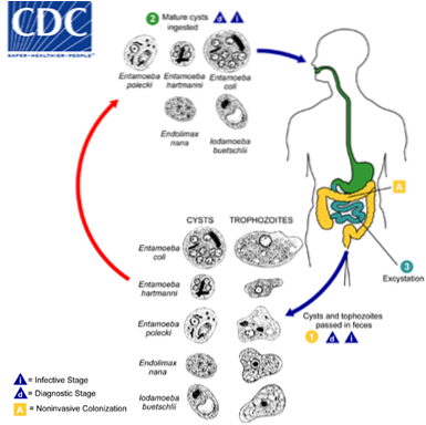
Similarly, phase 2 of excitation occurs in the small intestine, where phase 3 occurs, they are released and the trophozoites migrate to the large intestine. Thus, trophozoites replicate asexually producing cysts.
Due to the protection exerted by the configuration in their cell walls, the cysts survive for a few days or weeks outside the host organism, being responsible for the transmission.
Trophozoites that pass through feces are rapidly destroyed once they are outside the body, and if ingested they would not survive exposure to the gastric environment..
Stool culture is one of the most used techniques for diagnosis, although it can give false positives as it cannot differentiate from other species.
Other methods are tissue, genetic and molecular, in which biological products can be a biopsy, scraping an ulcer, blood, secretions from lesions, among others..
In this sense, determination through genetic and molecular evaluation is the most efficient to differentiate between pathogenic and non-pathogenic amoebas..
Entamoeba hartmanni, being a non-pathogenic amoeba, does not produce symptoms in carriers.
However, it has been found that under control conditions some non-pathogenic species manifest themselves to be associated with diarrheal diseases and symptoms..
This is not the case of E. hartmanni due to the great absence of research focused on it, so it is recommended that if symptoms are present, other analyzes should be carried out to determine their true origin..
The fact that it is a non-pathogenic amoeba avoids commenting on the treatment. Although, it is possible to find in the literature the use of Metronidazole and Tinidazole.


Yet No Comments