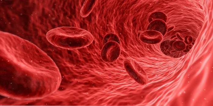
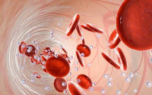
The erythrocytes, Also called red blood cells or red blood cells, they are very flexible and abundant blood cells, shaped like a biconcave disc. They are responsible for transporting oxygen to all body tissues thanks to the presence of hemoglobin inside the cell, as well as contributing to the transport of carbon dioxide and the buffering capacity of the blood..
In mammals, the interior of the erythrocyte basically consists of hemoglobin, since it has lost all subcellular compartments, including the nucleus. ATP generation is restricted to anaerobic metabolism.

Erythrocytes correspond to almost 99% of the formed elements present in the blood, while the remaining 1% is made up of leukocytes and platelets or thrombocytes. There are approximately 5.4 million red blood cells in one milliliter of blood..
These cells are produced in the bone marrow and can live an average of 120 days, in which they can travel more than 11,000 kilometers through the blood vessels.
Red blood cells were one of the first elements observed under the light of the microscope in the year 1723. However, it was not until 1865 that the researcher Hoppe Seyler discovered the oxygen transport capacity of said cell.
Article index
They are discoidal cells with an approximate diameter of 7.5 to 8.7 um and 1.7 to 2.2 um in thickness. They are thinner in the center of the cell than at the edges, giving a lifesaver appearance. They contain more than 250 million hemoglobin molecules inside..
Erythrocytes are cells with remarkable flexibility, since they must move during circulation through very thin vessels, about 2 to 3 um in diameter. When passing through these channels, the cell deforms and at the end of the passage it returns to its original shape.
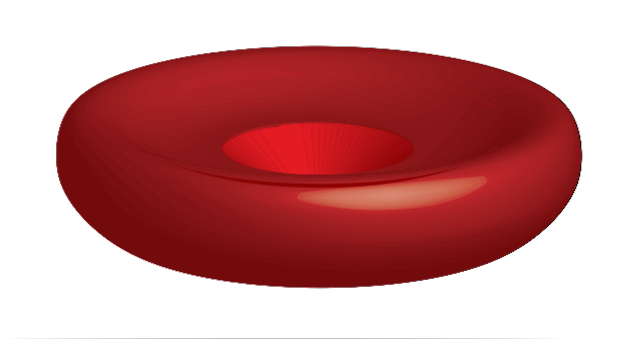
The cytosol of this structure contains the hemoglobin molecules, responsible for the transport of gases during blood circulation. The volume of the cell cytosol is around 94 um3.
When mature, mammalian erythrocytes lack a cell nucleus, mitochondria, and other cytoplasmic organelles, making them unable to synthesize lipids, proteins, or perform oxidative phosphorylation..
In other words, erythrocytes basically consist of a membrane that encloses the hemoglobin molecules..
It is proposed that erythrocytes seek to get rid of any subcellular compartment in order to ensure the maximum possible space for the transport of hemoglobin - in the same way that we would seek to remove all the elements from our car if we were to transport a large number of things.
The erythrocyte cell membrane comprises a lipid bilayer and a spectrin network, which together with the cytoskeleton, provides elasticity and compliance to this structure. More than 50% of the composition are proteins, slightly less lipids and the remaining portion corresponds to carbohydrates.
The erythrocyte membrane is the biological membrane that has received the most attention and of which there is the greatest knowledge, probably due to its ease of isolation and relative simplicity..
The membrane contains a series of integral and peripheral proteins connected to the lipid bilayer and spectrin. The connections that involve protein binding are known as vertical interactions and those that involve a two-dimensional array of spectrin by means of actin molecules are horizontal interactions..
When any of these vertical or horizontal interactions fail, it results in possible changes in spectrin density, in turn causing changes in erythrocyte morphology..
The aging of red blood cells is reflected in the stability of the membrane, reducing its ability to accommodate in the circulatory system. When this occurs, the monocyte-macrophage system recognizes the poorly functional element, eliminating it from circulation and recycling its content..
Proteins found in the cell membrane of erythrocytes can be easily separated on an electrophoresis gel. In this system, the following bands stand out: spectrin, ankyrin, band 3, proteins 4.1 and 4.2, the ion channel, glucophorins and the enzyme glyceraldehyde-3-phosphate-dehydrogenase..
These proteins can be grouped into four groups according to their function: membrane transporters, adhesion molecules and receptors, enzymes and proteins that bind the membrane with the components of the cytoskeleton..
The transporter proteins cross the membrane several times and the most important of this group is band 3, an anion exchanger of chloride and bicarbonate..
Because the erythrocyte is devoid of mitochondria, most enzymes anchor to the plasma membrane, including the glycolysis enzymes fructose-bisphosphate aldolase A, α-enolase, ALDOC, glyceraldehyde-3-phosphate dehydrogenase, phosglycerate kinase, and pyruvate kinase. kinase.
Regarding the structural proteins, the most abundant are band 3, spectrins, ankyrin, actin and band 4.1 protein, while band 4.2 protein, dematin, adduccins, tropomodulin and tropomyosin are considered minor components of the membrane.
Spectrin is a filamentous protein made up of an alpha and beta chain, whose structures are alpha helices.
The spectrin fibers resemble the springs of a mattress, and the portions of fabric that surround the mattress would represent the plasma membrane in this hypothetical example..
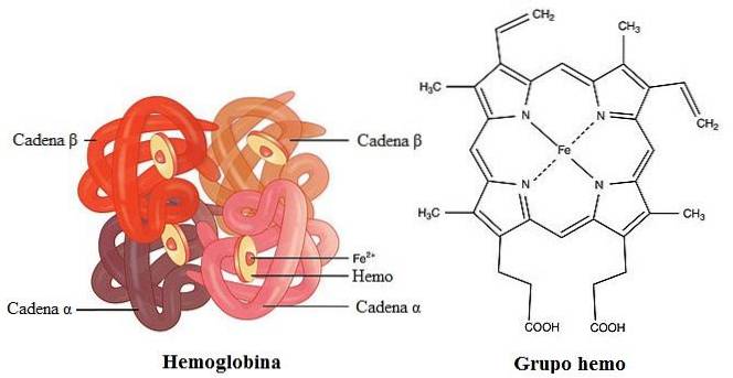
Hemoglobin is a complex protein with a quaternary structure synthesized in erythrocytes and is the fundamental element of these cells. It is made up of two pairs of chains, two alpha and two non-alpha (they can be beta, gamma or delta) linked together by covalent bonds. Each unit features a heme group.
It contains the heme group in its structure and is responsible for the characteristic red color of blood. Regarding its size, it has a molecular weight of 64,000 g / mol.
In adult individuals, hemoglobin is made up of two alpha and two beta chains, while a small portion replaces the beta with delta. In contrast, fetal hemoglobin is made up of two alpha and two gamma chains..
The oxygen that is diluted in the blood plasma is not enough to meet the demanding demands of the cell, for this reason it must exist in the entity in charge of transporting it. Hemoglobin is a protein molecule and is the oxygen transporter par excellence.
The most important function of erythrocytes is to house hemoglobin inside them to ensure the supply of oxygen to all tissues and organs of the body, thanks to the transport and exchange of oxygen and carbon dioxide. The mentioned process does not require energy expenditure.
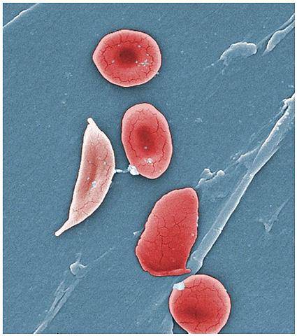
Sickle cell anemia or sickle cell anemia consists of a series of pathologies that affect hemoglobin, causing a change in the shape of red blood cells. The cells decrease their half-life time, from 120 days to 20 or 10.
The pathology occurs due to a unique change of an amino acid residue, glutamate for valine, in the beta chain of this protein. The condition can be expressed in its homozygous or heterozygous state.
The affected red blood cells take the shape of a sickle or a coma. In the image, normal blood cells are compared with pathological ones. In addition, they lose their characteristic flexibility, so they can break when trying to cross blood vessels..
This condition increases intracellular viscosity, affecting the passage of affected red blood cells through the smaller blood vessels. This phenomenon results in a decrease in the speed of blood flow..
Wound spherocytosis is a congenital disorder that involves the membrane of red blood cells. Patients who suffer from it are characterized by having a smaller diameter in the erythrocytes and a hemoglobin concentration greater than normal. Of all the diseases that affect the red blood cell membrane, this is the most common.
It is caused by a defect in the proteins that vertically connect the cytoskeletal proteins to the membrane. Mutations related to this disorder are found in the genes that code for alpha and beta spectrin, ankyrin, band 3, and proteins 4.2..
Affected individuals often belong to Caucasian or Japanese populations. The severity of this condition depends on the degree of the loss of connection in the spectrine network..
Hereditary elliptocytosis is a pathology that involves different changes in the shape of the erythrocyte, including elliptical, oval or elongated cells. This leads to a reduction in the elasticity and durability of red blood cells..
The incidence of the disease is from 0.03% to 0.05% in the United States and has been increased in African countries, since it provides some protection against the parasites that cause malaria, Plasmodium falciparum Y Plasmodium vivax. This same resistance is seen in individuals with sickle cell disease..
The mutations that produce this disease involve the genes that code for alpha and beta spectrin and protein 4.2. Thus, mutations in alpha spectrin affect the formation of alpha and beta heterodimer..
Hematocrit is the quantitative measure that expresses the volume of erythrocytes in relation to the total blood volume. The normal value of this parameter varies according to sex: in adult men it is 40.7% to 50.3%, while in women the normal range ranges from 36.1% to 44.3%.
In terms of cell number, in men the normal range is 4.7 to 6.1 million cells per uL, and in women between 4.2 and 5.4 million cells per uL.
Regarding normal hemoglobin values, in men it is between 13.8 to 17.2 g / dL and in women from 12.1 to 15.1 g / dL.
Similarly, normal values vary according to the age of the individual, neonates present hemoglobin values of 19 g / dL and gradually decreases until reaching 12.5 g / dL. When the child is young and is still breastfeeding, the expected level is from 11 to 14 g / dL.
In adolescent boys, puberty leads to an increase from 14 g / dL to 18 g / dL. For developing girls, menstruation can lead to a decrease in iron.
When the red cell count is less than the normal values mentioned above, it can be due to a number of heterogeneous conditions. The drop in red blood cells is associated with fatigue, tachycardia, and dyspnea. Symptoms also include paleness, headaches and chest pains.
The medical pathologies associated with the decline are diseases of the heart and of the circulatory system in general. Also pathologies such as cancer translate into low erythrocyte values. Myelosuppression and pancytopenia decrease blood cell production
Likewise, anemias and thalassemias cause a decrease in these blood cells. Anemias can be caused by genetic factors (such as sickle cell anemia) or by deficiency of vitamin B12, folate, or iron. Some pregnant women may experience symptoms of anemia.
Finally, excessive bleeding, whether from a wound, hemorrhoids, heavy menstrual bleeding or stomach ulcers, lead to loss of red blood cells..
The causes of high erythrocyte levels are just as diverse as those associated with low levels. The condition of exhibiting a high number of red blood cells is called polycythemia.
The most harmless occurs in individuals living in high regions, where the oxygen concentration is significantly lower. Also dehydration, in general, produces the concentration of red blood cells.
Diseases related to the kidneys, the respiratory system and cardiovascular diseases may be the cause of the increase.
Some external agents and harmful habits, such as smoking, can increase the red blood cell count. Long-term use of cigarettes lowers blood oxygen levels, increasing demand and forcing the body to generate more erythrocytes.
The consumption of anabolic steroids can stimulate the production of red blood cells in the bone marrow, as can doping with erythropoietin, which is used to optimize physical performance..
In some cases of anemia, when the patient is dehydrated, the plasma-lowering effect counteracts the decrease in red blood cells, resulting in a deceptively normal value. The pathology comes to light when the patient is hydrated and abnormally low erythrocyte values can be evidenced.


Yet No Comments