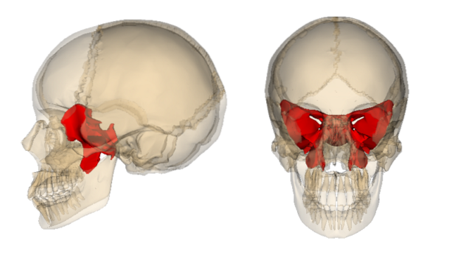
The sphenoid It is an odd bone of the skull that is part of the facial skeleton. It is located in the middle part of the skull, behind the frontal bone and the ethmoid and in front of the occiput. It is one of the seven bones that articulate to form the orbit.
It is shaped like a butterfly or bat, since it has a central body with lateral wings. In its structure, it has multiple orifices and channels through which neurological and vascular structures open..
In its lower portion, it has a projection on each side called the pterygoid process, which serves as an insertion surface for several of the muscles of the face. Multiple neurological elements run through this process.
The body of the sphenoid is hollow and forms the so-called sphenoid sinus, one of the eight sinuses. These air cavities of the bones are structures that influence phonation, in regulating the temperature of the air that enters through the nose and as a defense in infectious processes, among other functions..
Due to the relationships of the sphenoid with important nerves and arteries of the face and skull, its injuries involve serious sequelae for the patient, so they must be treated in a timely manner..
Article index
The sphenoid begins its formation from the 8he week of gestation in a complex process in which its body is formed first with the notch for the pituitary gland and later its wings. By that time, these elements are separated.
Around 9to week the cartilaginous ossification nuclei begin to form that will eventually unite the bone into a single structure.
The sphenoid sinus, which is the hollow portion of your body, is formed from the 12to week, when a cartilaginous portion invades the back of the bone and forms a cavity that will fill with air years after birth.
The origin of the sphenoid is parallel to that of the brain, so it can be associated with some rare birth defects, such as transsphenoidal encephalocele which is the exit of part of the brain through the body cavity of the sphenoid, due to abnormalities in its formation.
The sphenoid bone is one of the 22 bones that make up the skull and one of the 8 that make up the orbit. Represents the boundary between the neurocranium and the facial skeleton, joining both structures.
It is a large, complex bone that occupies the middle part, below the base of the skull. In front it limits with the frontal bone and with the ethmoid, and behind with the occiput. Its anterior limits allow stability to the skull and make an adequate and strong cavity for the brain.
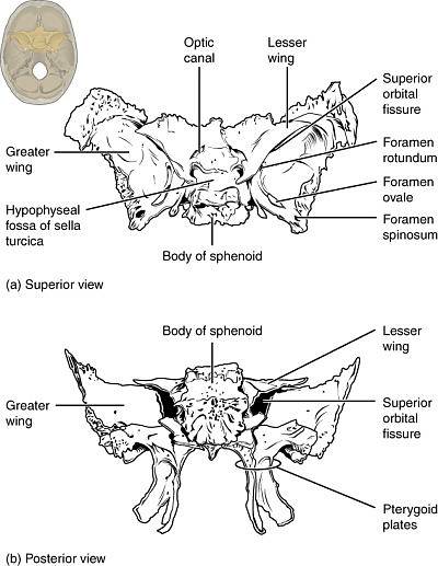
It is made up of a cuboid body and lateral structures called sphenoid wings, in which two portions are recognized: major and minor.
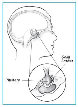
In the body of the sphenoid a depression called turkish saddle, this is where the pituitary gland is located. This body is hollow and forms one of the eight paranasal sinuses, the so-called sphenoid sinus.
The sphenoid has multiple orifices and passage channels through which important vascular and neurological structures run. There are the optic canal, for the optic nerve, the foramen ovale, the superior orbital fissure and the spinous foramen.
In the position it occupies, it is articulated with 12 bones. Unique four: vomer, ethmoid, frontal and occipital; and 6 pairs: temporal, zygomatic, parietal and palatal.
The pterygoid process is a protrusion of the sphenoid that is located on each side of the point where the body meets the greater wing.
It is pyramidal in shape with a lower vertex and upper base. Two blades are described in its structure, one lateral and one medial..
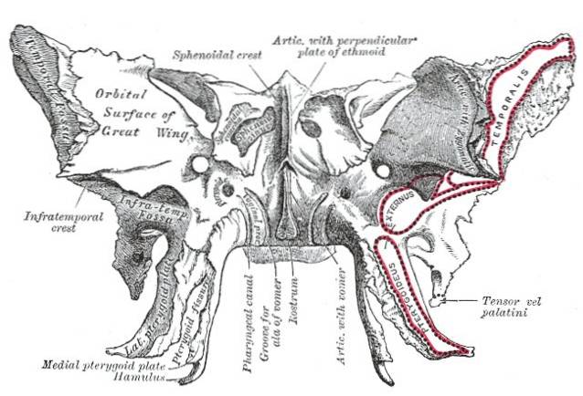
The medial one is horseshoe-shaped, its inner edge serves as an insertion surface for the tendon of the tensor palati muscle, while its outer edge constitutes part of the lateral limit of the choanas, which are the inner openings of the nasal cavity..
On the other hand, the lateral pterygoid and median pterygoid muscles are inserted in the lateral lamina. Together with the temporal bone it contributes to the formation of some orifices for the passage of neurological structures.
The sphenoid bone is essential at the junction of the facial and cranial bones. Its relationship and articulation with the rest of the bone structures, gives rigidity to the skull.
It also serves as an insertion surface for various muscles, especially the pterygoid process, where the chewing muscles are inserted..
It acts as protection for the important vascular and neurological structures that have passage between the brain, the facial space and the cervical.
The sphenoid sinus, like the rest of the paranasal sinuses, helps reduce the weight of the skull, drain nasal secretions, warm the air that enters the nose, protect against respiratory infections and improve resonance during phonation..
Sphenoid fractures are complex and serious injuries that must be diagnosed and treated in a timely manner..
Partial or complete loss of vision is a common complication in injuries to the orbital portion of the bone. Thus, there may be multiple neurological sequelae depending on the degree of injury, due to the multiple nerves that cross the bone..
The appearance of some signs such as that of Battle, which is the hematoma in the cutaneous projection of the mastoid process, may indicate injury to the sphenoid bone.
Whenever there is suspicion of a skull base fracture with cranial nerve dysfunction, the possibility of injury to the sphenoid bone should be investigated..
The fracture of the pterygoid process falls into the group of fractures of the midface called LeFort fractures.
Any facial fracture that involves severe trauma to the nose or frontal bone may involve the pterygoid process and the sphenoid bone..
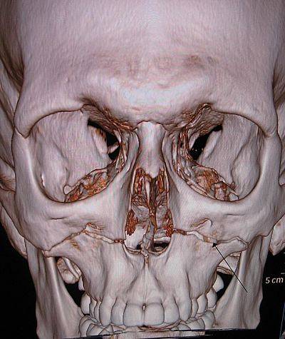
They are diagnosed from the history and physical examination. In turn, confirmation is made by imaging studies such as plain skull radiography and computerized axial tomography (CT)..
The treatment of these fractures is surgical, since it is a life-threatening injury that affects the stability of the skull..


Yet No Comments