The sphingomyelin it is the most abundant sphingolipid in animal tissues: its presence has been verified in all cell membranes studied to date. It has structural similarities with phosphatidylcholine in terms of the polar head group, which is why it is also classified as a phospholipid (phosphosphingolipid).
In the 1880s, scientist Johann Thudichum isolated an ether-soluble lipid component from brain tissue and named it sphingomyelin. Later, in 1927, the structure of this sphingolipid was reported as N-acyl-sphingosine-1-phosphocholine.
Like the other sphingolipids, sphingomyelin has both structural and cell signaling functions, and is especially abundant in nervous tissues, specifically in myelin, a sheath that covers and isolates the axons of certain neurons.
Its distribution has been studied through subcellular fractionation and enzymatic degradation experiments with sphingomyelinases, and the results indicate that more than half of the sphingomyelin in eukaryotic cells is found in the plasma membrane. However, this depends on the cell type. In fibroblasts, for example, it accounts for almost 90% of total lipids.
The dysregulation of the synthesis and metabolism processes of this lipid lead to the development of complex pathologies or lipidosis. An example of these is hereditary Niemann-Pick disease, characterized by hepatosplenomegaly and progressive neurological dysfunction.
Article index
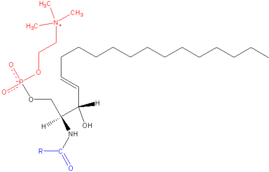
Sphingomyelin is an amphipathic molecule composed of a polar head and two apolar tails. The polar head group is a phosphocholine molecule, so it may appear similar to the glycerophospholipid phosphatidylcholine (PC). However, there are substantial differences regarding the interfacial and hydrophobic region between these two molecules..
The most common base in a mammalian sphingomyelin molecule is ceramide, composed of sphingosine (1,3-dihydroxy-2-amino-4-octadecene), which has a double bond in trans between the carbons at positions 4 and 5 of the hydrocarbon chain. Its saturated derivative, sphinganine, is also common, but is found to a lesser extent.
The length of the hydrophobic tails of sphingomyelin ranges from 16 to 24 carbon atoms and the fatty acid composition varies depending on the tissue.
The sphingomyelins of the white matter of the human brain, for example, possess nervonic acid, those of the gray matter contain mainly stearic acid, and the prevalent form in platelets is arachidonate..
There is generally a disparity in length between the two fatty acid chains of sphingomyelin, which seems to favor "interdigitation" phenomena between hydrocarbons in opposite monolayers. This provides the membrane with special stability and particular properties compared to other membranes that are poorer in this sphingolipid..
In the interfacial region of the molecule, sphingomyelin has an amide group and a free hydroxyl at carbon 3, which can serve as donors and acceptors of hydrogen bonds for intra- and intermolecular bonds, important in the definition of side domains and interaction with various types of molecules.
The products of sphingosine metabolism -ceramide, sphingosine, sphingosine 1-phosphate and diacylglycerol- are important cellular effectors and give it a role in multiple cellular functions, such as apoptosis, development and aging, cell signaling, among others..
Thanks to the three-dimensional “cylindrical” structure of sphingomyelin, this lipid can form more compact and ordered membrane domains, which has important functional implications from the protein point of view, since it can establish specific domains for some integral membrane proteins.
Lipid rafts, membrane phases or ordered micro domains of sphingolipids such as sphingomyelin, some glycerophospholipids and cholesterol, represent stable platforms for the association of membrane proteins with various functions (receptors, transporters, etc.).
Caveolae are invaginations of the plasma membrane that recruit proteins with GPI anchors and are also rich in sphingomyelin.
Cholesterol, due to its structural rigidity, significantly affects the structure of cell membranes, especially in aspects related to fluidity, which is why it is considered an essential element.
Because sphingomyelins possess both hydrogen bond donors and acceptors, it is believed that they are capable of forming more “stable” interactions with cholesterol molecules. For this reason, it is said that there is a positive correlation between the levels of cholesterol and sphingomyelin in the membranes..
The synthesis of sphingomyelin occurs in the Golgi complex, where the ceramide transported from the endoplasmic reticulum (ER) is modified by the transfer of a phosphocholine molecule from phosphatidylcholine, with the concomitant release of a diacylglycerol molecule. The reaction is catalyzed by SM synthase (ceramide: phosphatidylcholine phosphocholine transferase).
There is also another pathway of sphingomyelin production that can occur by transferring a phosphoethanolamine from phosphatidylethanolamine (PE) to ceramide, with subsequent phosphoethanolamine methylation. This is thought to be particularly important in some PE-rich nerve tissues..
Sphingomyelin synthase is found on the luminal side of the Golgi complex membrane, which is consistent with the extra cytoplasmic location of sphingomyelin in most cells.
Due to the characteristics of the polar group of sphingomyelin and the apparent absence of specific translocases, the topological orientation of this lipid depends on the enzyme synthase..
Sphingomyelin degradation can occur in both the plasma membrane and lysosomes. Lysosomal hydrolysis to ceramide and phosphocholine depends on acidic sphingomyelinase, a soluble lysosomal glycoprotein whose activity has an optimal pH of around 4.5.
Hydrolysis in the plasma membrane is catalyzed by a sphingomyelinase that works at pH 7.4 and that requires divalent magnesium or manganese ions for its operation. Other enzymes involved in the metabolism and recycling of sphingomyelin are found in different organelles that connect to each other through vesicular transport pathways..


Yet No Comments