
The sperm They are the mature sex cells (gametic cells) produced in the male gonads. They are highly specialized cells, fully dedicated to the task of fertilizing female ovules, a fundamental event during sexual reproduction..
They were discovered more than 300 years ago by Antony van Leeuwenhoek, who, motivated solely by his curiosity, observed his own semen and coined the term “animalculus” to the flagellated structures that he observed..
Since then, these cells have been the object of study of many investigations, especially those related to fertility and assisted reproduction.
Sperm are cells with high energy requirements, as they must move at high speed once they are ejaculated from the penis (male reproductive organ) to the vaginal tract (female reproductive organ).
The energy they use derives mainly from the metabolism of carbohydrates such as glucose, that is, from glycolysis and mitochondrial oxidative phosphorylation, which was demonstrated in 1928, thanks to the experiments carried out by McCarthy et al..
The formation and release of these cells depends on many endocrine (hormonal) factors, especially testosterone, which is produced and secreted by the testes..
Unlike what happens with female sex cells (which are produced during embryonic development), sperm are produced continuously throughout the adult life of man..
Article index
Sperm are very important cells, since they have the special task of fusing with the ovum contained in the female ovaries to fertilize and fertilize it, a process that ends with the formation of a new individual..
Sperm, as well as ovules, are haploid cells, so the fusion of the female and male nuclei restores the diploid charge (2n) in a new cell. This implies that each cell contributes half of the chromosomal load of a human being in this process..
In humans, sperm are the cells responsible for determining the sex of the progeny, since the ovum has an X sex chromosome, but each sperm can have either an X chromosome or a Y chromosome..
When the sperm that successfully fertilizes and fertilizes the egg has an X chromosome, the baby that will be formed will be XX, that is, it will be genetically female. On the other hand, when the sperm that fuses with the egg has a Y chromosome, the baby will be XY, that is, genetically male.
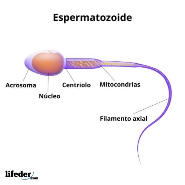
Sperm are small flagellate cells (less than 70 microns in length). Each sperm is made up of two well-defined regions known as the head and tail, both enclosed by the same plasma membrane..
In the head is the nucleus that will serve to fertilize the female ovum, meanwhile the tail is the organelle of locomotion that allows them to move and that represents an important part of their length.
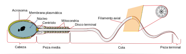
The sperm head is flattened in shape and is roughly 5 microns in diameter. Inside it is the cellular DNA, which is very compacted, which minimizes the volume it occupies, facilitating its transport, transcription and silencing..
The sperm nucleus has 23 haploid chromosomes (in a single copy). These chromosomes differ from the chromosomes of somatic cells (cells in the body that are not sex cells) in that they are packed with proteins known as protamines and some sperm histones..
Protamines are proteins with abundant positive charges, which facilitate their interaction with negatively charged DNA.
In addition to the nucleus, the head of the sperm has a secretory vesicle known as the acrosomal vesicle or the acrosome, which partially surrounds the anterior region of the nucleus and is in contact with the plasma membrane of the sex cell..
This vesicle houses a large number of enzymes that facilitate the process of penetration of the outer covering of the ovum during fertilization. These enzymes include neuraminidase, hyaluronidase, acid phosphatase, arylsulfatase and acrosin, a protease similar to trypsin..
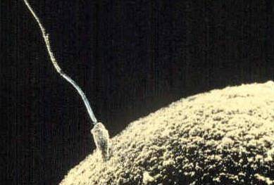
When the egg and the sperm come into contact with each other, the acrosome releases its contents by exocytosis, a process known as the “acrosome reaction”, essential for the union, penetration and fusion of the sperm with the ovum.

The head and tail of the sperm are covered by the same plasma membrane. The tail is a very long flagellum that has four regions called the neck, middle piece, main piece and end piece..
The axoneme, that is, the cytoskeletal structure that provides movement to the tail, emerges from a basal body located behind the nucleus of the sperm. This basal body is what makes up the neck and is about 5μm long..
Between the neck and the end piece is the intermediate piece. It is 5 microns long and is characterized by the presence of multiple mitochondria that are arranged in the form of a "sheath" around the central axoneme. These highly specialized mitochondria are what provide, in essence, the energy necessary for movement in the form of ATP..
The main piece is just under 50 μm long and is the longest part of the tail. It begins in a "ring" that prevents further advancement of the mitochondria and ends in the end piece. As you get closer to the end piece, the main piece tapers (tapers).
The terminal piece, finally, is made up of the last 5 μm of the tail and is a structure where a certain “disorder” is observed in the microtubules that make up the axoneme of the flagellum..
An average adult man produces millions of sperm per day, however these cells take 2-3 months to fully form and mature (until they are ejaculated).
The life cycle of a sperm cell begins with gametogenesis or spermatogenesis, that is, with the division of a germ or precursor cell, which gives rise to cell lines that divide later, to later differentiate and mature. In the meantime, defective cells undergo programmed cell death processes.
Once formed in the seminiferous tubules, the maturing sperm must migrate into a region of the testis known as the epididymis, which is approximately 20 feet long. This migration takes a few days and it has been shown that at this stage the cells are not mature enough to fertilize an egg, as they lack sufficient mobility.
After 18 or 24 hours have passed in the epididymis, the sperm are perfectly mobile, but this mobility is inhibited by certain protein factors.
Once in the epididymis, the sperm maintain their fertility for little more than a month, but this time will depend on the conditions of temperature, diet and lifestyle..
When sperm are ejaculated during intercourse (sexual intercourse), they have full capacity for movement, moving at speeds as fast as 4 mm / min. These cells can survive for 1 to 2 days in the female reproductive tract, but this depends on the acidity of the surrounding environment..
Sperm production (spermatogenesis) first occurs in humans during puberty. This process takes place in the testes, which are two organs of the male reproductive system, and has to do with the reduction of the chromosomal load of the sex cells (which go from being diploid (2n) to being haploid (n)).
In the testes, spermatogenesis occurs within ducts known as seminiferous tubules, the epithelium of which is made up of two main types of cells: Sertoli cells and spermatogenous cells..
The spermatogenous cells give rise to the spermatozoa, while the Sertoli cells nourish and protect the spermatogenous cells. The latter are in the seminiferous tubules in different stages of maturation.
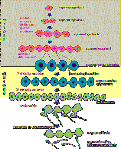
Among the spermatogenous cells are cells known as spermatogonia, which are immature germ cells responsible for dividing and producing primary spermatocytes, secondary spermatocytes, and mature sperm.
Spermatogonia are located towards the outer edge of the seminiferous tubules, near their basal lamina; as they divide, the cells they give rise to migrate towards the central portion of the ducts, where they finally mature.
Spermatogonia divide by mitosis (asexual division) and are diploid cells (2n) that when dividing generate more spermatogonia and primary spermatocytes, which are nothing more than spermatogonia that stop dividing by mitosis to enter meiosis I.
A small group of spermatogonia slowly divide by mitosis throughout life, functioning as “stem cells” for the mitotic production of more spermatogonia or cells that are committed to maturation..
When spermatogonia mature, that is, when they divide by mitosis and later by meiosis, their progeny do not complete the cytosolic division, so the daughter cells (clones) remain connected to each other by cytosolic bridges, as if they were a syncytium..
This syncytium is maintained until the final stages of maturation and migration of sperm cells (sperm), where the sperm is released into the lumen of the seminiferous tubules. This results in groups of cells being produced synchronously..
Primary spermatocytes, as they divide by meiosis, form secondary spermatocytes, which divide again by meiosis (meiosis II), differentiating themselves into another type of cell called spermatids, which have half the chromosomal load of spermatogonia. say, they are haploid.
When spermatids mature, they differentiate into mature spermatozoa thanks to a series of morphological changes that involve the elimination of a large part of their cytosol, the formation of the flagella and the internal rearrangement of their cytosolic organelles..
Some of these changes have to do with the condensation of the cell nucleus, with the elongation of the cell and the rearrangement of the mitochondria.
These cells subsequently migrate to the epididymis, a kinky tube in the testes, where they are stored and continue the maturation process. However, only through a process known as capacitation, which takes place in the female genital tract, do sperm complete their maturation..


Yet No Comments