
The stereocilia they are specializations of the outer and apical surface of the plasma membrane of some epithelial cells. They are immobile and very rigid microvilli that form branched brush-like “tufts”.
Stereocilia are found in the cells of the epididymis (the organ located at the posterior border of the testis, where sperm mature and are stored) and in the piliform cells or sensory cells of the cochlea, in the inner ear.
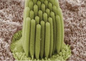
They are long fingerlike processes of the apical portion of the plasma membrane of these cells. They measure 100 to 150 nm in diameter and are about 120 μm long at most. When looking at a group of stereocilia you can see branched fingerings of different lengths.
They are composed of actin, which is a protein that makes up the cell cytoskeleton. Actin is bound to other fibrin filaments and to the plasma membrane through ezrin, another protein. The separation between one stereocilium and another is approximately 10 nm.
In the epididymis, stereocilia increase the surface area of the membrane and fulfill functions of absorption and secretion of a liquid that constitutes one of the components of semen..
In the sensory cells of the inner ear, these structures fulfill functions related to the generation of signals, that is, they participate in the process of mechano-transduction (transformation of a mechanical signal into an electrical signal)..
Article index
The distinguishing feature of stereocilia is their rigidity. Unlike the other specializations of the surface of the plasma membrane, these fingerings do not have their own mobility and although they increase the surface area of the membrane, they have specialized functions.
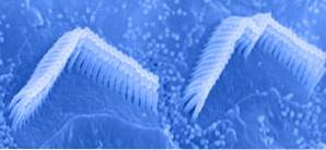
In the inner ear, specifically in the mammalian cochlea, the stereocilia are arranged in an orderly and symmetrical manner. Each row is made up of stereocilia of the same size, in such a way that the stereocilia of the parallel rows form a "descending ramp".
In the cochlea, these stereocilia are bathed in endolymph, a fluid that bathes the membranous labyrinth of the inner ear with an ionic composition similar to that of intracellular fluid. That is, it has a high concentration of K + and a low concentration of Na+.
Due to these characteristics of endolymph, the sensory cells of the inner ear have very different electrophysiological characteristics from other cells in the body. While most cells are excited by the entry of sodium, they do so by the entry of potassium.
This particularity is the cause of the temporary deafness that accompanies the use of some medications called diuretics, which increase urinary volume. Some diuretics increase urinary losses of K + and the decrease in this ion causes deafness.
The structure of stereocilia is very simple. They have a central portion with actin, which gives them rigidity. In turn, actin binds to fibrin fibers and ezrin, which binds it to the plasma membrane.
In the mammalian cochlea, each hair cell is provided with 30 to a few hundred stereocilia arranged in three rows of different sizes and symmetrically and bilaterally. One row of long stereocilia, one medium and one row of shorter stereocilia on each side of the cochlea.
Each stereocilium, at its insertion site in the membrane, becomes sharper and ends up forming a kind of hinge on which it pivots or rotates. These basal movements of the hinge area are related to the opening of channels and the transformation of a mechanical movement into an electrical signal..
In the cochlea, each stereocilium has an ion channel at its luminal end. This channel is a protein that forms a pore whose opening is regulated by a gate. The gate is connected to a regulating “spring”, sensitive to tension or stretching..
Each spring is connected to the spring of the higher neighboring stereocilium by means of very fine elastic extensions. These extensions are called "point joints" or "end connections".
The upper part of the stereocilia remains rigid thanks to its embedding in the reticular lamina (for those that belong to the inner cells) and in the tectorial membrane (for those that belong to the outer cells).
These two membranes (tectorial and the reticular lamina) undergo sliding movements of one over the other in the same direction, but on different axes, for which the stereocilia embedded in them bends due to shear movements..
In the epididymis, the stereocilia fulfill some secretory functions very different from those of the cochlea, however they are structurally similar.
The function of the stereocilia of the sensory cells of the inner ear is to provoke a receptor potential that induces the release of neurotransmitters in the nerve fiber connected to it (which is directed to the central nervous system) and originates a generator potential.
This occurs due to the mechanical deformation suffered by the stereocilia due to the movement of the endolymph..
The endolymph moves as a consequence of the transmission of sound waves through the eardrum and the movement of the chain of ossicles of the middle ear.
As the movement of the stereocilia towards the higher stereocilia occurs, the tension generated at the junctions opens the gate of the cation channel and K + and Ca ++ enter the sensory cell. This excites the cell, generating an electrical depolarization called "receptor potential." This initiates the release of neurotransmitters in the basal part of the cell that synapses with the afferent fiber..
The main neurotransmitter released is excitatory and produces a generator potential in the nerve fiber that, upon reaching the threshold, causes an action potential.
The action potential in the primary nerve fibers, in turn, initiates the stimulation of the nerve pathway that ends in the areas of the brain responsible for hearing. In this way we perceive the sound.
The function of the stereocilia of the epididymis is related to the reabsorption of part of the fluid that enters the epididymis from the testes. In addition, they contribute to the secretion of a liquid known as "ependymal liquor" which is part of the liquid components of semen..


Yet No Comments