The stomachache or stomodeum It is an ectodermal depression that appears around the fourth week of embryonic development and, initially, is the center of the development of facial structures. Derived from the Greek stoma- (mouth) and odaios- (similar to) that means "it looks like a mouth".
This depression is located between what will be the skull and the pericardium of the embryo, forming part of the foregut. It is the precursor of the mouth and the anterior lobe of the pituitary gland (adenohypophysis). Initially it constitutes the oral and nasal cavity together, since there is still no separation between the two.
The stomodeum is lined with ectoderm and separated from the anterior end of the foregut by the oropharyngeal membrane. This membrane disappears at the end of the third week of intrauterine development or fifth week of embryonic development, thus establishing oropharyngeal communication..
By the fourth and a half weeks of embryonic development, the stomodeum shows a series of mesenchymal elevations. These elevations are the caudal mandibular processes, the maxillary processes, located laterally, and the single, rounded frontal prominence in a cranial or superior direction..
Ectoderm thickenings appear on each side of the frontal prominence and immediately above the stomodeum, giving rise to what is known as the "nasal placode", which will participate in the formation of the nostrils..
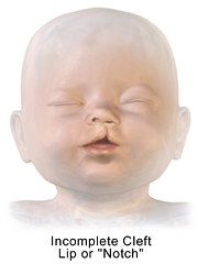
Congenital malformations in this area can affect the palate, lips and nostrils. Several are the resulting alterations among which the cleft lip and cleft palate can be named..
Article index
Due to the bending or cephalocaudal folding of the embryo, the brain or cranial structure approaches the pericardial cavity, leaving a depression or cleft between both structures that is called the stomodeus..
Thus formed, the stomodeum is initially delimited or blocked in the posterior part by a membrane that separates it from the foregut in its cephalic portion. Laterally, in the upper part, is the encephalic prominence, on the floor is the pericardium of the embryo and forward it opens towards what will be the amniotic cavity.
As the embryo bends the stomodeus and the primitive intestine are delineated. Subsequently, the oropharyngeal membrane ruptures and leaves the stomodeum in communication with the upper part of the anterior or pharyngeal intestine, a structure that will give rise to the pharynx.
Between the fourth and fifth week of embryo development, the stomodeus presents a series of elevations or prominences formed by the proliferation of mesenchyme. This shows the maxillary processes laterally, the mandibular processes caudally and the frontal prominence cranially..
Once the palate and the upper and lower jaws have developed, the stomodeus becomes the oral cavity, which is now separated from the nasal cavity.
As already explained previously, the stomodeus is formed by the bending of the embryo that leaves the cleft between the cephalic portion and the pericardial region of the same.
Initially, the stomodeum constitutes the nasal and oral cavity together, open forwards (towards what will be the amniotic cavity) and closed backwards by the oropharyngeal membrane, which separates them from the pharyngeal intestine or foregut (which is a portion of the so-called intestine primitive).

The different elements that develop from the mesenchymal proliferations that develop in the walls of the stomodeum will give rise to most of the facial structures..
Thus, the mandibular processes or processes will form the lower jaw or maxilla. The maxillary processes located laterally on both sides of the stomodeus grow in an internal direction and end up merging with each other and laterally with the mandibular processes, thus forming the cheeks and delimiting the size of the oral cavity.
In the frontal prominence appears the nasal placode from which the nasolateral and nasomedial processes will develop around it. These processes will form the nostrils, the wings of the nose, the middle portions of the nose, the upper lip and the maxilla, as well as the entire primary palate..
The pituitary gland develops in two completely different portions: the first is an ectodermal evagination of the stomodeum that develops just in front of the oropharyngeal membrane, called Rathke's pouch; the second is the infundibulum, a downward extension of the diencephalon.
In the three-week embryo, Rathke's bursa is a prominence within the stomodeum in its postero-superior part and it grows dorsally towards the infundibulum. After the second month, it is no longer observed within the oral cavity and is very close to the infundibulum.
Later, as the development continues, the cells in the anterior part of this bag grow rapidly and form the anterior lobe of the pituitary or adenohypophysis. The infundibulum will give rise to the posterior pituitary or neurohypophysis. Cells at the back of the bursa cause the pars intermediate of the gland.
The function of the stomodeum is to be the center of the embryonic development of the facial structures and the anterior part of the pituitary called the adenohypophysis..
Within the facial structures that develop, the stomach cavity will form the oral cavity and the lateral structures the other components already listed. The mouth is a fundamental part of the digestive system, since the initial part of the digestive process occurs in it..
Some elements such as teeth, tongue and glands have other origins, but they develop parallel to the development of the oral cavity. For example, the parotid and submandibular glands appear in the connective tissue of the cheek as it develops..
Around the 10th week of development, the face has already formed. Note the nose with well developed nasolacrimal folds and nostrils.
The upper lip grooves are found and both the upper and lower lips are well shaped and fused. The maxilla, mandible, and palate have already developed, and the eyes and pinna are seen. The oral cavity has already formed with its internal structures already formed.


Yet No Comments