The tertiary structure of proteins It is the three-dimensional conformation that polypeptide chains acquire when they fold on themselves. This conformation appears by interactions between the side chains of the amino acid residues of the polypeptide. Side chains can interact regardless of their position in the protein.
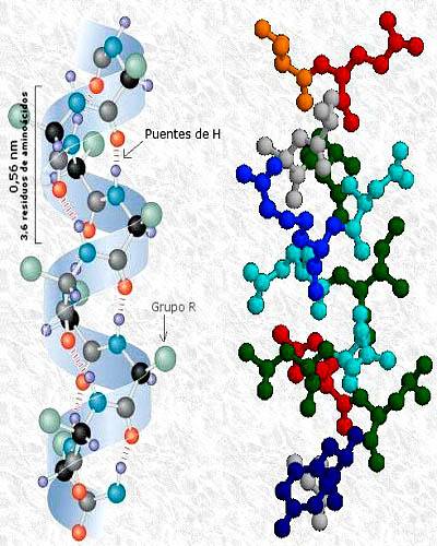
Because it depends on the interactions between the R groups, the tertiary structure shows non-repetitive aspects of the chain, since these groups are different for each amino acid residue. The secondary structure, on the other hand, depends on the carboxyl and amino groups, which are present in all amino acids.
Some authors suggest that fibrous proteins have a simple tertiary structure, but nevertheless, other authors point out that this structure is typical of globular proteins..
Article index
In fibrous proteins, the polypeptide chains are arranged in the form of long filaments or long sheets; they are generally made up of a single type of secondary structure. This secondary structure is, in most cases, more important than the tertiary structure in determining the shape of the protein..
Its biological function is structural, giving strength and / or elasticity to the organs and structures where they are found, while keeping them together. All fibrous proteins are insoluble in water, due to the large amount of hydrophobic amino acid residues they present.
These fibrous proteins include keratins and collagen. The former are found in connective tissues and in structures such as hairs, nails (α-keratins), scales and feathers (β-keratins). Collagen, for its part, is found in bones, tendons and skin, among others.
These proteins are part of the so-called intermediate filament proteins, which play an important role in the cytoskeleton of multicellular organisms. In addition, they are the main constituent of hair, nails, wool, horns, hooves, and one of the main proteins of animal skin..
The structure of the molecule is an α helix. Two α-keratin strands can be arranged in parallel and wound one over the other with their hydrophobic R groups interacting with each other. In this way, a superhelical structure or ball with winding to the left is created..
The tertiary structure of α-keratin is simple and is dominated by the secondary structure of α-helix. On the other hand, the quaternary structure is also present, since two molecules participate in the superhelical structure, which interact through non-covalent bonds..
The primary structure is similar to that of α-keratins, but their secondary structure is dominated by β sheets. They are the main constituent of reptile scales and bird feathers..
This protein can represent more than 30% of the total protein mass of some animals. Found in cartilage, bones, tendons, the cornea, and skin, among other tissues.
The secondary structure of collagen is unique, being represented by a left-handed helix with 3.3 amino acid residues for each turn. Three left-handed helix chains (α-chains) wrap around each other giving a right-handed supercoiled molecule, known by some authors as tropocollagen..
Tropocollagen molecules come together to form a collagen fiber that has a high strength, superior to that of steel and comparable to that of high-strength copper.
Other types of fibrous proteins are fibroin and elastin. The first one is made up of β sheets, consisting mainly of glycine, alanine and serine..
The side chains of these amino acids are small in size, so they can be tightly packed. The result is a fiber that is both very resistant and very little extensible..
In elastin, for its part, valine replaces serine among its main constituent amino acids. Unlike fibroin, elastin is very extensible, hence its name. In the constitution of the molecule, lysine also acts, which can participate in crosslinks that allow elastin to regain its shape when tension ceases.
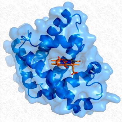
Globular proteins, unlike fibrous ones, are soluble and generally have several types of secondary structures. However, in these, the three-dimensional conformations that they acquire when folding on themselves are more important (tertiary structure).
These particular three-dimensional conformations confer specific biological activity on each protein. The main function of these proteins is regulatory, as with enzymes.
The tertiary structure of globular proteins has some important characteristics:
- Globular proteins are compact thanks to packing by folding the polypeptide chain.
- The distant amino acid residues in the primary structure of the polypeptide chains remain close together, being able to interact with each other due to folding..
- Larger globular proteins (more than 200 amino acids) can have several compact segments, independent of each other and with particular functions, and each of these segments is called a domain. A domain can have between 50 and 350 amino acid residues.
As already pointed out, proteins present particular forms of folding, which also give them particular characteristics. This folding is not random and is favored both by the primary and secondary structure and by some non-covalent interactions, and there are also some physical restrictions to the folding, for which some rules have been formulated:
- All globular proteins have defined distribution patterns, with the hydrophobic R groups directed towards the interior of the molecule and the hydrophilic residues in the outer layer. This requires at least two layers of secondary structure. The β-α-β loop and the α-α vertex can provide these two layers.
- The β-sheets are generally arranged in a left-handed rolled form..
- In a polypeptide chain, different turns can occur to pass from one secondary structure to another, such as β or γ turns, which can reverse the direction of the chain by four amino acid residues or less..
- Globular proteins possess α-helices, β-sheets, turns, and irregularly structured segments.
If a protein loses its native (natural) three-dimensional structure, it loses its biological activity and most of its specific properties. This process is known by the name of denaturation.
Denaturation can occur when natural environmental conditions change, for example by varying temperature or pH. The process is irreversible in many proteins; however, others may spontaneously regain their natural structure when normal environmental conditions are restored..

Yet No Comments