The Euplots They are a genus of ciliated protozoa that move freely on the surface of muddy waters, from where they obtain the bacteria necessary for their food.
These microorganisms are called ciliates because they have the presence of cilia, hair-like appendages, essential for their movement from one place to another and for obtaining food..
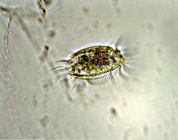
The cilia it presents are grouped in tufts called cirrus clouds, which the microorganism uses as a paddle or to walk, depending on the surface where it is. These cirrus clouds are in front, on the sides and at the end of its body, resembling a tail..
The ventral area (belly) of these organisms is flat and the dorsal area (back) is bulky or ribbed, resembling a coffee bean. It has several separate ribs that run the length of the body from end to end.
Most of the current ciliates correspond to the species Euplotes Charon They have an oval shape and transparent appearance. They live in areas of slow or stagnant water circulation.
Article index
The body of the Euplotes is made up of: ectoplasm, contractile vacuole (mouth), cirri, membranelas, neuromotor apparatus, anal opening, endoplasm, macronucleus and micronucleus.
Its body is transparent, rigid, oval, measures approximately 80 to 200 µm long and is distinguished by a macronucleus that is visible inside, in the shape of an inverted “C”, with an adjacent micronucleus..
The mouth of the Euplotes is in the anterior region and its perimeter is triangular. This mouth is large and has cilia around it, which form a membrane that resembles fangs. When these cilia move, they allow them to eat diatom algae and small particles of plant material..
Despite this challenging appearance, they are calm, harmless and peaceful beings, unlike the Paramecians, who have a harmless appearance but are really dangerous..
On the side, the Euplotes look quite thin and you can see their cilia joined in tufts to form the cirrus, which it uses to move around. Sometimes they have a ciliary row on each side of the ventral area.
The cirri located in the lateral and rear areas have a spiny appearance and allow the mobility of these microorganisms, to climb or walk, other times to swim according to the need and the environment..
The number and location of ventral cirrus clouds in Euplotes, and the geometry of the ventral argyrome, are the criteria used to divide this taxon into four morphologically different subgenera: Euplotes, Euplotoides, Euplotopsis and Monoeuplotes..
Taxonomically, Euplotes are classified as follows: Biota Chromista (Kingdom) Harosa (Sub-kingdom) Alveolata (Infra-kingdom) Protozoa (Phylum) Ciliophora (Sub-phylum) Ciliata (class) Euciliata (Sub-class) Spirotricha (Order).
In turn, within the genus Euplotes, there are the following species
Euplotes aberrans, Euplotes acanthodus, Euplotes aediculatus, Euplotes affinis, Euplotes alatus, Euplotes antarcticus, Euplotes apsheronicus, Euplotes arenularum, Euplotes balteatus, Euplotes balticus, Euplotes affinis, Euplotes alatus, Euplotes antarcticus, cristalusrassus crabs , Euplotes euryhalinus, Euplotes eurystomus, Euplotes focardii, Euplotes gracilis, Euplotes harpa, Euplotes iliffei, Euplotes latus, Euplotes mediterraneus, Euplotes minor, Euplotes minuta, Euplotes moebupiusiotes, Euplotes nectopolitans, Euplotes musculature parabalteatus, Euplotes parawoodruffi, Euplotes patella, Euplotes poljanski, Euplotes quinquecincarinatus, Euplotes quinquicarinatus, Euplotes raikovi, Euplotes rariseta, Euplotes salina, Euplotes sinica, Euplotes strelkovius, Euplotesonewitch zuplotesoneuplotesonewitchonus, Euplotesone strelkovius, Euplotesonewitchonwitch, Euplotesone strelkovius.
It is common to observe Euplotes in both fresh and salty waters. When used for microbiological experimentation and other cellular analysis techniques, they should be preserved in mixed cultures with molds, algae, yeasts, bacteria or other protozoa that serve as food..
Under these conditions, laboratory work options for biochemical tests, for example, are limited. But due to its large size and diversity of organizational patterns, its experimental use remains a great advantage over the technical deficiencies of cultivation..
These particular ciliates are easy to collect due to their ubiquity (they are found anywhere in the world) and can be grown comfortably in the laboratory, making them a great tool for studying biological processes in general..
In natural environments, Euplotes must deal with predators. This prey-predator interaction forces them to use two types of defense: individual and group.
In the individual escape strategy, the microorganism is capable of reacting and moving away from predators that carry out toxic discharges in radii of 300 microns in diameter and in a maximum time of 90 seconds..
The group escape strategy is more refined and complex. These ciliates have a low concentration non-protein molecule that generates a repulsive action to repel predators. A few Euplotes from each demographic group are qualified to secrete such a substance that encourages the escape of predators..
Euplotes have a very wide bioecological range and are considered cosmopolitan species, due to their physiological diversity that gives them great adaptability.
They can be located in different ecosystems such as the coastal waters of California, Japan, Denmark and Italy. It is also common to locate them in plankton such as benthic ciliates and there are also some that colonize snow particles.
The diet of the Euplotes is very varied and they use several feeding tactics. They consume cells of different sizes, from bacteria to diatom algae and also eat other protozoa.
They can be omnivorous, consume bodontids (a type of flagellates) and a great variety of heterotrophic flagellates (which transform organic matter into nutrients and energy), including other classes of ciliates..
Some species have selective feeding, such as Euplotes vannus. Some studies describe a relationship between the type of food, its concentration and the growth of the population of these microorganisms.
The reproduction of Euplotes is especially characteristic due to the DNA synthesis process that occurs in the macronucleus..
In some species, such as Euplotes eurystomus, the reproductive generation time is short and its growth is high, if the medium where it is found is adequate. This species uses Aerobacter aerogenes as its main source of food..
Most protozoa reproduce asexually, by mitotic cell division, but some species have the ability to reproduce sexually, through a process called: conjugation.
When Euplots mate, there is an exchange of genetic material through a bridge of cytoplasm. After this exchange, the new generation that has been formed by cell division will make various combinations of genes from the cells of the parents..
After fertilization, the cells separate when the diffusion zone is reabsorbed and the contraction processes become operative. Many specialists consider that the sexual cycle is superimposed on an asexual cycle that precedes it.
Sometimes a mating called intraclonal conjugation or selfing occurs and occurs when there is no sexual or asexual fertilization..
This is advantageous because it restores the life cycle clock and disadvantageous because it can only be carried out for a short time since it can lead to a loss of adaptation due to loss of genetic variation..


Yet No Comments