The stool examination It is a laboratory study in which fecal matter (feces) is examined in order to detect intestinal parasites. It is one of the simplest and oldest laboratory techniques, being initially developed by Anton Van Leeuwenhoek in the 18th century..
Anton Van Leeuwenhoek, considered the father of microbiology, used the "direct" coproparasitic-toscopic method to observe his own feces and described what years later were identified as the trophozoites of Giardia lamblia, a protozoan that invades the small intestine of man.
Parasitic diseases affect millions of people in the world, especially in poor or underdeveloped countries, where there are poor sanitary conditions related to the disposal of excreta and the consumption of contaminated water..
The diagnosis of these diseases is important for an adequate treatment, being the coproparasitoscopic examination is an indispensable tool for it. It is a simple, fast and inexpensive laboratory test.
The stool examination includes several techniques that, in addition to allowing direct visualization and quantification of eggs, trophozoites, cysts, or larvae, allows identifying the structures of the microorganism and thus identifying the parasite..
Techniques used for stool examination include methylene blue staining techniques, concentration methods, Faust, Richie technique, sedimentation techniques, and direct, single, or serial examinations..
Article index
To carry out this study, the patient is required to take a fresh stool sample that is not contaminated with urine, water, blood (menstrual) or soil. The sample must be the size of a walnut or, if it is liquid, it must be at least the volume corresponding to two tablespoons.
The patient must not have taken parasiticidal drugs for at least three days prior to taking the sample or for the period indicated by their doctor. You should also not use laxative medications.
Samples should be placed in a dry, wide-mouth, lidded container or a specially designed disposable container (available at your preferred pharmacy). Samples should be placed in a cool environment, should not be refrigerated for more than 24 hours, and should not be stored near heat sources or frozen..
When the indicated examination is serial, at least three samples are required, which must be taken every 24 hours or more, as indicated by the physician. For these cases, laboratories generally provide a set of bottles containing solutions with preservatives..
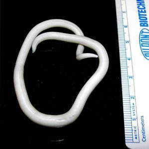
When the patient observes a “worm” in the stool, if possible, he should place it in a closed bottle with water and take it to the laboratory together with the bottle of the stool sample..
The vials with the samples or with the "worms" must be labeled and identified with the name of the patient, his age, sex and the date and time of the sample collection..
It is very important to adequately instruct the patient on all these aspects of taking and handling the samples, since it depends on whether the elements that may exist in said sample remain viable for observation, identification and diagnosis..
There are direct stool tests and sample suspension and concentration techniques that are used many times to avoid false negatives and observe much cleaner samples. Some staining techniques are also used to identify certain parasites..
The direct stool examination, by the technique of drop earring, consists of making a dilution of the stool sample with physiological solution (0.9% NaCl) and placing a drop of this solution in a concavity that has a special slide used for this purpose.
Once the drop is placed on the slide, it is covered with a coverslip and observed under the microscope. This technique allows us to observe eggs and cysts, but it also allows us to observe any mobile element such as flagellates, larvae, trophozoites, ciliates, etc..
Suspension techniques use a solution that is denser than the elements to be observed, so that these float on the surface of the liquid and can be collected, since they remain concentrated in the surface layer of the solution..
This technique has the advantage that it allows to have a fairly clean sample of detritus, since these, being more dense, remain at the bottom of the bottle. The relative disadvantage is that the solution shrinks and deforms the microorganisms in a short time.
These methods are not used for helminth and cestode eggs because they are very heavy and do not float in these solutions. They are widely used to observe protozoa in their tropozoic form or their eggs and for the observation of larvae such as those of Strongyloides stercoralis.
Another technique widely used for not deforming the microorganisms in the sample and being simple and economical is the formalin sedimentation technique.
Among the concentration techniques we can cite as an example the techniques of Faust and Richie.
The different techniques that allow the microscopic visualization of the eggs, larvae or other elements of the different intestinal parasites, combined with the staining techniques, allow the identification and diagnosis of these diseases..
Next, a clinical case is described and some images are shown that illustrate the usefulness of the stool examination for the diagnosis and the evaluation of the benefits of the treatment..
An 18-year-old male patient comes to the doctor's office for colicky abdominal pain, more intense in the periumbilical area, nausea and episodes of watery diarrhea.
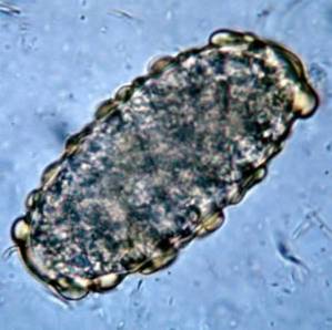
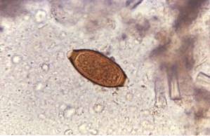
When questioning the patient, the doctor notes two salient points: 1) the patient refers to having bathed in a lake in a rural area and 2) he is struck by the fact that his feces float in the toilet. After examining the patient, the doctor suspects the presence of Giardia lamblia.
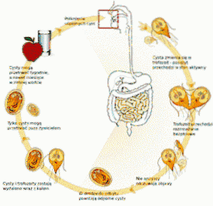
This protozoan is lodged in the small intestine of man and interferes with the absorption of fats, which generates very greasy stools that tend to float. Pollution frequently occurs from polluted waters in lakes or streams in rural areas or in pools or hot tubs with poor maintenance.
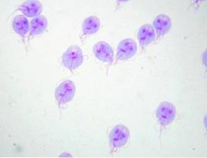
The doctor indicates a stool examination and the results confirm the presence of Giardia lamblia. After the end of the treatment, another stool examination is indicated that confirms the absence of cysts or trophozoites of Giardia lamblia.


Yet No Comments