Fasciola hepatica is a worm that belongs to the phylum of flatworms, specifically to the class Trematoda. Also known by the name of fluke, it has been studied in depth, since it is responsible for a disease known as fascioliasis, which mainly affects the liver and gallbladder tissues.
It was described for the first time by the famous Swedish naturalist Carlos Linnaeus in 1758. This is a parasite that has a very particular life cycle, in which there is an intermediate host (snail) and a definitive host (mammals like humans)..
Preventive measures against the disease caused by this worm include avoiding the consumption of aquatic plants in regions where the parasite is common..
Article index
This is a parasite that belongs to the Eukarya domain. As such, it is made up of eukaryotic-like cells. This means that each and every one of your cells has a cellular organelle known as the nucleus..
Inside this is the genetic material (DNA) forming the chromosomes. In this same order of ideas, Fasciola hepatica It is considered multicellular, because it is made up of various types of cells.
Fasciola hepatica it is an organism that, from the embryonic point of view, is triblastic. This implies that it presents the three germ layers: endoderm, ectoderm and mesoderm. From them the various organs that make up the animal are formed.
They also do not have coelom, so they belong to the group of acellomed animals. In regards to symmetry, Fasciola hepatica It has bilateral symmetry, since it is made up of two exactly equal halves.
On its reproduction, in the life cycle of Fasciola hepatica it is appreciated that it has both asexual and sexual reproduction. The latter occurs within its definitive host, while asexual reproduction occurs within the intermediate host (snail)..
Fasciola hepatica It is an animal that presents indirect development because throughout its life it must go through various larval stages. It is also oviparous because it reproduces through eggs..
The taxonomic classification of Fasciola hepatica is the next:
-Domain: Eukarya
-Animalia Kingdom
-Phylum: Plathyhelminthes
-Class: Trematoda
-Subclass: Digenea
-Order: Echinostomida
-Family: Fasciolidae
-Gender: Fasciola
-Species: Fasciola hepatica
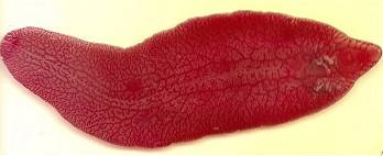
Fasciola hepatica it is an unsegmented worm that is shaped like a flattened leaf. Adult individuals are approximately 3.5 cm long by 1.5 cm wide. It has a cephalic and a ventral zone.
In each of these areas you can see suction cups through which they can attach themselves to their guests. The suction cup in the cephalic area is smaller than the one in the ventral part.
The body of the parasite is covered by an integument, which has a large number of folds and spines that the parasite uses to optimize its absorption process.
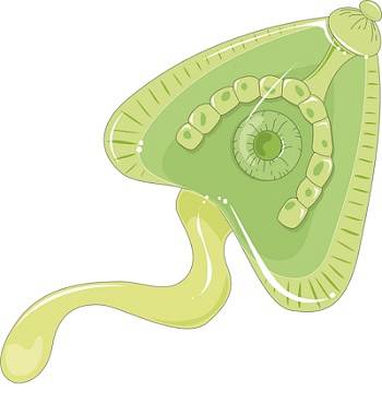
The internal morphology of the parasite is quite simple. Its digestive system is basic and incomplete, since it does not have an anus. It is made up of the mouth opening that opens into a cavity, which continues with a pharynx and the esophagus. The latter divides and ends in structures called the intestinal cecum..
The nervous system is made up of neuronal clusters or ganglia. While its excretory system is of the protonephridial type.
Fasciola hepatica It is a hermaphroditic animal, which implies that it has reproductive organs, both male and female. His testicles, two in number, are branched. The ovary is in the right half of the animal and the uterus is short.
The life cycle of Fasciola hepatica It is a bit complex, since it includes several stages and two hosts, an intermediate one (freshwater snail) and a definitive one, which is generally a mammal such as cattle. In many cases, the definitive host is the human being.
The cycle begins inside the definitive host, specifically at the level of the bile ducts, which is where the adult parasite fixes. In this place, the parasite lays the eggs, which are carried through the intestine, together with the fecal material to the outside.
Those eggs that are released are not embryonated. This means that the embryo does not begin to develop until the egg leaves the external environment. Here, it develops into a larva known as the miracidium. This larva manages to leave the egg thanks to the action of certain digestive enzymes that disintegrate the operculum of this.
The miracidium is a larva that is characterized by presenting cilia and having the ability to move freely in the aquatic environment. It should be noted that it is the infectious form of this parasite for its intermediate host.
As already mentioned, the intermediate host of Fasciola hepatica is a freshwater snail, generally those of the species Limnaea viatrix. It is important to mention that miracidium takes approximately 8 hours to find a snail, since it cannot survive in the environment for longer..
Once it locates a host, the miracidium is located at the level of the snail's foot and slowly pierces its cells in order to enter its interior. There the miracidia undergo a change and transform into sporocysts.
Sporocysts go through a process of asexual reproduction known as parthenogenesis, through which they give rise to the next stage known as redias. Finally the redias are transformed into cercariae, which end up leaving the body of the snail.
This larval stage (cercariae) have the ability to move freely through the water for an approximate period of time of about 10 hours. At the end of these, they lose their tail and generally adhere to aquatic plants, encysting themselves, transforming into metacercariae. The latter constitutes the infectious form for the definitive hosts (mammals)..
When metacercariae are ingested by mammals such as cows, goats, sheep, and even man, they travel through the digestive tract until they reach the intestine. Specifically in its first portion (duodenum), they cross the intestinal wall and lodge in the peritoneal cavity for approximately two weeks..
Later, they are able to move towards the liver. There, already converted into immature flukes, they feed on the liver tissue for about 8 weeks. After this time, when they have reached maturity, they move to their final place of confinement: the bile ducts..
There in the bile ducts they cause damage and havoc and feed on the blood that is produced in the injuries it generates. It is at this site that sexual reproduction occurs that results in the formation and release of eggs..
Fasciola hepatica it is a heterotrophic organism because it cannot synthesize its own nutrients, but must feed on other living beings or substances produced by them. In this sense, it belongs to the group of hematophages.
A blood-sucking animal is one that feeds on the blood of other animals. In the particular case of Fasciola hepatica, it attaches itself to the bile duct with the help of its suction cups, perforates the blood vessels and feeds on the host's blood.
Fasciola hepatica it is a pathogenic organism that generates in mammals that are its definitive hosts, a disease known as fascioliasis.
This disease has three variants: acute, chronic and latent. In addition to this, in the course of the disease two stages or phases are distinguished: the initial one, which covers from the moment the host ingests the metacercariae, until the parasite fixes itself in the bile ducts..
The second stage is known as state. In this, the parasite becomes sexually mature and begins to release eggs in the host's feces..
The symptoms that manifest in fascioliasis are varied, although most are limited to the organs affected by the parasite as it moves through the host's body until it reaches its definitive place..
The acute phase of the disease is the initial one. In it the symptoms are given by the damage caused by the parasite in the peritoneal cavity and when they reach the liver. Consider the following symptoms:
-Elevated body temperature (Fever)
-Hepatomegaly (Enlargement of the liver)
-Eosinophilia (Increased eosinophils in the blood)
-Severe abdominal pain
-General discomfort
-Weightloss
-Digestive symptoms such as nausea and vomiting (rare symptoms).
When the disease is not treated in time, it becomes chronic. The signs and symptoms that appear in this stage are the following:
-Jaundice from liver and biliary damage
-Pancreatitis
-Abdominal pain that can be diffuse and intermittent
-Cholelithiasis
-Cholangitis
-Biliary cirrhosis.
Infection by Fasciola hepatica can be diagnosed through direct methods and indirect methods.
These methods are based on the identification of eggs from Fasciola hepatica in the patient's stool or in the bile. The fact that the test is negative does not necessarily exclude infection with this parasite. This is because the eggs are produced when the parasite has already reached sexual maturity..
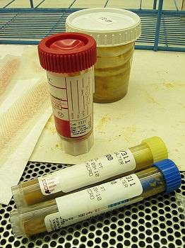
Due to this, it is important that a serial examination is carried out, using different types of dyes, such as lugol or eosin..
Indirect methods are not related to the direct detection of the parasite, but to the identification of the antibodies that the host generates and that circulate throughout its bloodstream. The technique through which this test is performed is ELISA (enzyme linked immunosorbent assay).
To perform this test, there must be a clear suspicion of an infection by Fasciola hepatica, based on the clinical manifestations of this. This must be so because this is not a routine exam and it also involves a significant investment of money.
It is important to note that the examination that clearly demonstrates the presence of this parasite in the host is the identification of its eggs in the stool examined..
Taking into account that Fasciola hepatica it is a parasite, the medicines used to treat its infection are anthelmintics. The drug generally chosen by specialist doctors is triclabendazole.
This medicine acts at the level of the parasite's metabolism, preventing it from using glucose for its energy processes. Because of this, the parasite ends up dying.
Sometimes nitazoxanide can also be used.


Yet No Comments