The reticular fibers They are fine strands of connective tissue that form a network that supports the tissue of many organs. The name of reticular fiber is due to its organization in a pattern similar to that of a mesh or network.
Reticular fibers, together with collagen fibers and elastic fibers, make up the extracellular matrix. This matrix is an intricate and complex structural network that surrounds and supports cells in connective tissue..
Fibroblasts are the main cells of connective tissue. They are responsible for the synthesis of reticular, collagen and elastic fibers, and carbohydrates.
Article index
Reticular fibers are synthesized by fibroblasts called reticular cells. They are composed of type III collagen.
They are thin, with a diameter less than 2 µm. They exhibit periodicity with D pattern bands, similar to collagen fibers, although diametrically they are thinner and more uniform. They form a network by branching and anastomosis with other reticular fibers.
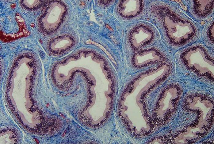
Using a light microscope, reticular fibers cannot be visualized when hematoxylin and eosin are used to stain tissues. They are specifically dyed, acquiring a black coloration, by impregnation with silver. This differentiates them from type I collagen fibers, which acquire a brown color..
The presence of carbohydrates in the reticular fibers gives them a high affinity for silver. For this reason it is said that the reticular fibers are argentofílicas.
The distribution of reticular fibers is rather restricted. They are found in the basement of epithelial tissue, the surface of adipose cells, muscle cells, Schwann cells, the sinusoid endothelium of the liver, and lymphoid tissue. The prevalence of reticular fibers is an indicator of tissue maturity.
Reticular fibers differ in structure, organization, and function from collagen fibers. Both types of fiber form an extensive and continuous network of collagen fibrils.
Beneath the basal lamina, the reticular fibers form a delicate network of thin fibrils. The individual fibrils are firmly attached to the basal lamina, forming a distinctive structural unit that demarcates and supports the cellular components of different tissues and organs..
In the lymph nodes there is a structural skeleton formed by a reticular network consisting of elastin and reticular fibers. This skeleton supports the lymphatic vessels and sinuses within the tissues. The organization of the reticular fibers provides a space for the movement of molecules in the extracellular fluid..
Reticular fibers are prominent in the initial stages of tissue healing, where they represent an early extension mechanism of the extracellular matrix, which is newly synthesized.
Type III collagen of reticular fibers plays a role in the extensibility of embryonic tissue, in which they are prominent. During embryonic development, reticular fibers are replaced by type I collagen fibers, which are stronger.
Lymph nodes are secondary lymphoid organs with a highly organized and compartmentalized structure.
Lymph nodes provide: 1) a system of "highways" that facilitate lymphocyte migration; 2) an environment that favors interactions between different types of cells of the immune system; 3) a system for sending mediators to critical sites.
These functions depend on a reticular cell network, which consists of reticular fibers associated with the extracellular matrix and reticular cells. The membranes of these cells form an envelope in the center of which are collagen fibers, where they form the extracellular matrix..
The fibers are woven throughout the lymph node. Many of these fibers traverse the sinus of the nodule, continue through the superficial cortex between the follicles, and penetrate a dense network of the deep cortex..
The reticular cell network is important for the immune response. Small molecules, coming from surrounding tissue or from pathogens, such as protein fragments, can be distributed by reticular fibers.
Some viral infections damage the reticular network of cells. For example, diphtheria toxin destroys reticular cells. Lymph nodes tolerate the loss of up to half of their reticular cells.
The network of reticular fibers of the pancreas forms an interstitial compartment, through which the capillaries pass. It completely occupies the space between the constituents of the parenchyma of the gland. This shows that this interstitial compartment serves for the passage of fluid from the capillaries..
The islets of Langerhans of the pancreas are surrounded by a capsule of reticular fibers, which has the function of maintaining the cells as a functional unit.
Within the islet, the reticular fibers are found around the capillaries and form a three-dimensional sheath. The thin layer of reticular fibers separates the islets from the exocrine tissue of the pancreas.
During embryo formation, hematopoiesis takes place at different sites in the body, including the liver, spleen, lymph nodes, and bone marrow. After birth, hematopoiesis takes place exclusively in the bone marrow.
In the bone marrow there is a loose organization of thin reticular fibers, which form an intricate connective tissue network. In adulthood, the bone marrow is confined to the bones of the skull, sternum, ribs, vertebrae, and pelvic bones..
In these bones, the stroma of the connective tissue is made up of reticular cells and reticular fibers that form a delicate meshwork, which surrounds the islands of hematopoietic cells and provides support to the bone marrow..
Ehler-Danlos syndrome type IV is the result of an error in the transcription of DNA or in the translation of the messenger RNA that codes for type III collagen, which is the main component of reticular fibers..
Symptoms are thin, translucent, and fragile skin that is easily injured and abnormally flexible. Patients may present with a ruptured intestine and large arteries, in which reticular fibers surround smooth muscle cells..


Yet No Comments