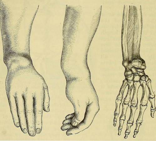
The Colles fracture It is a total fracture of the radius, the forearm bone, at its distal end. This injury is distinguished by a displacement of the fractured portion backwards, which produces abnormal angulation and deformity in the limb. It is the most common type of bill of the forearm bones.
Bone injury commonly occurs from trauma that causes a linear, transverse rupture of the end of the bone. The relationship with the wrist and the action of the associated muscles causes the characteristic dorsal mobilization. The mobilization of the displaced bone fragment is estimated to be about 30 mm dorsally.
The Irish surgeon Abraham Colles was the one who first described the injury in 1814. The doctor in his observations describes both the posterior displacement of the fractured segment and the deformation of the limb; In honor of these observations, the name Colles fracture was born..
The radius is one of the long bones of the forearm, located between the elbow and wrist joints. It is a long bone in the shape of a prism, slightly curved and occupies the external side of the limb. At its distal end, the cortex is usually thinner, which predisposes it to fracture more easily.
This type of fracture is more common in youth and from the sixth decade of life. It is more common in women than men, and is generally related to falls, work-related or sports accidents. It is rare to find growth cartilage injuries in children due to this type of injury..
The alteration of shape and disability resulting from Colles' fracture warrants immediate treatment. This treatment consists of returning the bone fragment to its original position, which may involve surgery. Medical significance is due to temporary or permanent disability for physical activity and work.
Article index
The mechanism of Colles fracture is a trauma that occurs when the outstretched hand receives the impact of trauma.
Usually this occurs after a fall and the defensive response of stopping with the hand. The causes will depend on the age, activity carried out and clinical conditions of the patient..
Colles' fracture occurs most often in children, adolescents, and the elderly. In the former it occurs due to their physical activities and games, in addition to the weakness of the bones in children.
In the elderly, the presence of osteoporosis and instability in locomotion make fractures due to falls more frequent.
Athletes, workers and drivers are more likely to have accidents.
Osteoporosis, vertigo, cerebrovascular disorders and cardiovascular diseases predispose to the occurrence of falls and fractures.
- Falls from your own feet.
- Falls from height.
- Car accidents.
- Accidents due to sports activities or the practice of extreme sports.
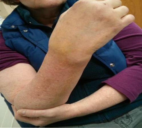
The symptoms in Colles' fracture are those associated with a long bone fracture: pain, soft tissue edema, deformity and functional limitation.
Other symptoms can appear as a consequence of complications. Once the reduction of the fracture and immobilization is carried out, the symptoms will disappear progressively.
Pain is the cardinal symptom that is present in trauma. In the case of bone fractures, pain occurs due to a rupture of the periosteum, the layer that covers the bone.
The periosteum has a large number of sensory fibers, so a bone lesion is capable of causing the pain that characterizes it..
Soft tissue trauma can trigger pain, due to stimulation of superficial sensory receptors and release of inflammation-producing substances.
As a consequence of the trauma, edema -increased volume- occurs in the soft tissues. This is due to the release of inflammatory mediators and increased interstitial fluid..
The bone marrow of the long bones is highly irrigated, and the fracture can cause bleeding and consequently localized bruising..
Loss of continuity of a bone causes deformity or loss of its anatomical configuration. In the case of Colles' fracture, the deformity is caused by the posterior displacement of the end of the broken radius. The resulting shape of the limb is called a fork, "s" or bayonet deformity, a clinical sign of this fracture..
Also called functional impotence. Radio-ulnar and radio-carpal joints allow free mobility of the hand.
Rupture of the distal end of the radius causes alteration of both joints, limiting the normal range of motion of the hand. In addition, the pain already described considerably impedes the function of the limb..
Paresthesia or sensory disturbances - tingling, burning, or cramps - may occur in the hand. Neurological symptoms are associated with injury to the median nerve or due to prolonged immobilization of the limb.
Vascular damage is rare. It is possible the presence of fractures in the ulna or bones of the wrist that worsen the symptoms.
Although it does not occur frequently, soft tissue injuries such as skin, ligaments, or tendons may accompany a radius fracture..
This would complicate the injury and lengthen the recovery time. Secondary infections can occur adding fever, redness and local heat to the symptoms..
Treatment of Colles' fracture is intended to restore the anatomy and function of the radius and its joints. Therapy includes general measures, reduction of the fracture, immobilization and subsequent rehabilitation.
Both reduction, immobilization and rehabilitation will be the responsibility of emergency physicians and specialists.
Anti-inflammatory analgesics are used to reduce pain. Antibiotics will be used in case of associated infections. Neurological symptoms, if present, are treated with B complex and antineuritics.
Local ice application reduces swelling and bruising.
Reduces pain and is a measure prior to consultation with the specialist.
Also called closed reduction. It consists of restoring the position of the radius by non-surgical measures. This procedure must be performed by qualified personnel, such as emergency physicians or orthopedists..
It is a conservative measure used in cases of minor angulations, and there is a risk of recurrence of the fracture.
It is an invasive surgical procedure that consists of reducing the focus of the fracture through surgery. Osteosynthesis material - plates, screws or surgical wire - is used to stabilize the already reduced fracture.
The reduction can be performed by external or internal fixation and the surgery will be performed exclusively by traumatologists.
After reduction of the fracture, the limb must remain immobilized by using plaster bandages (plaster). Rigid immobilization encompasses the distal third of the arm, forearm, and palm of the hand.
The plaster should be changed between 7 to 10 days after its placement since, by reducing the edema, it loses its immobilizing effect..
Once the fracture has been resolved and immobilization is removed, the rehabilitation phase follows. Both the fracture and prolonged immobilization produce some degree of muscle atrophy and shortening of the tendons.
The patient will be referred to a physiotherapy service to perform exercises that facilitate full functional recovery.


Yet No Comments