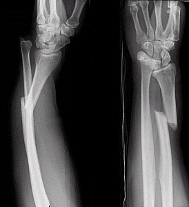
The Galeazzi fracture It is a fracture of the distal third of the radius that involves the distal radioulnar joint. It is caused by direct trauma to the wrist or by falling with the hand in hyperextension and with the forearm in pronation (movement of the forearm that allows the hand to be placed with the back up).
The Galeazzi fracture was first described by Sir Astley Cooper in 1822 and later, in 1934, it was named after the Italian surgeon at the Rachitti Institute in Milan, Riccardo Galeazzi. This surgeon presented 18 cases of this type of fracture.
It is a rare fracture in adults. It is more common in men than women, with a frequency of 3 to 7% among all wrist fractures. It is more common in children.
Symptoms associated with this fracture consist of pain in the wrist and forearm that is exacerbated by movement, regional hematoma, edema, soft tissue deformation, and a soft area on palpation of the fracture site..
It is associated with instability of the radio-ulnar joint; resolution of the fracture in adults requires surgical treatment, otherwise closed resolution is associated with recurrent dislocation of the distal radius joint.
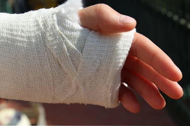
Most of the cases of these fractures in young children, after being reduced, can be treated with immobilization with a cast, without the need for surgical intervention..
Article index
Galeazzi fracture is a fracture of the lower third of the radius with injury or dislocation of the distal radioulnar joint.
Sometimes it involves a sector near the middle of the shaft of the radius and at other times it can also include an ulna fracture. In the latter case, this fracture is called “Galeazzi-like fracture”Or“ Galeazzi-type fracture ”.
When the fracture is less than 7.5 cm from the distal radius joint, 55% of patients present with joint instability. On the other hand, when the fracture occurs at a distance greater than 7.5 cm from the joint, only 6% of patients present instability of said joint.
They are fractures difficult to treat and when they are reduced by closed mechanisms and immobilized with a plaster, they are associated with sequelae and pathologies in the recovery process. The treatment of choice is surgical and should include resolution of the fracture and joint injury..
Numerous classifications have been reported for Galeazzi fractures, one of the last being published in 2014. However, the Association of Traumatology and Orthopedics ("Orthopedic Trauma Association", OTA) presents a classification called “OTA Classification” for Galeazzi fractures.
The latter classifies these fractures into three types: Type A, Type B and Type C. In turn, each type has several categories, as explained below:
1.1. Only the ulna with intact radius
1.2. Only the radius with intact ulna or ulna
1.3. Fracture of both bones
2.1. Only the ulna with intact radius
2.2. Only the radius with intact ulna
2.3. Fracture of both bones
3.1. Only the ulna with intact radius
3.2. Only the radius with intact ulna
3.3. Fracture of both bones
In children, diaphyseal radius and ulna fractures are one of the most frequent and can be complete, complete displaced, bun or green stem. These fractures can occur in the middle, distal, or proximal third of the shaft of the bone, although most occur in the distal third..
Children with these fractures, if not displaced or rotated, are treated orthopedically with cast immobilization for 6 to 8 weeks. If the fracture is displaced or rotated, it is reduced (sometimes under general anesthesia) and then a cast is placed for the same period.
Surgical solutions in children are exceptional, they are only indicated when there is a vascular or nervous complication. When required, a fasciotomy (cutting the fascia) may be done to relieve pressure that may be compressing a vessel or nerve and impede blood flow. These are also indicated in open fractures.
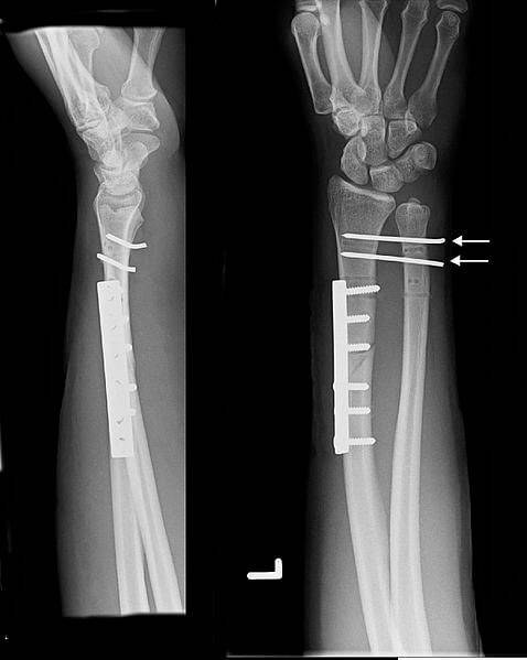
In adults, Galeazzi fractures have a surgical indication. There are three types of surgical treatments for these fractures: intramedullary nail placement, external fixation with stakes, or plate and screw fixation..
Of these three types of surgical treatments, plate fixation is the most frequently used for Galeazzi fracture, as it achieves early functional mobilization and stable, uncomplicated consolidation in 95% of cases..
To correct the joint injury, external fixation and immobilization systems are usually used for about 4 to 6 weeks, and then the fixation system is removed after 6 to 8 weeks.
On the one hand, the objective of rehabilitation is to promote the formation of bone callus (magnetotherapy is used for this) and, on the other hand, is to avoid complications and obtain the maximum possible functional level..
Among the complications that can be avoided are the atrophic effects of immobilization, inflammation and pain, stiffness of joints that remain immobile for a long time, among others..
Generally, while the cast or external fixation is in place, mobilization exercises are done for the shoulder joint on the affected side, avoiding the appearance of stiffness in these joints. Isometric exercises are used and mobilization exercises are also done for the fingers.
Once the immobilization period is over, progressive flexion and extension exercises are carried out for the wrist and elbow applying resistance. Pronosupination exercises are not indicated before the eighth week. Exercises for the entire upper limb are included to restore function after immobilization..
The most frequent complications are the following:
- The bone is fractured again once the plaque is removed.
- Persistent pain even after plaque removal.
- No bone union has occurred after treatment.
- That the consolidated union is defective.
- Infections.
- Neurological injuries.
- Radioulnar synostosis (fusion of both bones)


Yet No Comments