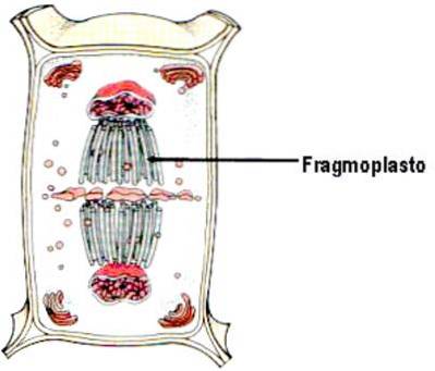
The fragmoplasts are structures formed mainly by a set of microtubules or microfibrils that are arranged in a barrel shape within the dividing plant cell and are formed during late anaphase (third phase of mitosis) or telophase (fourth and last phase of mitosis) early.
Cytokinesis is the final stage of the cell cycle and consists of the separation and segmentation of the cytoplasm. This process takes place during the last phase of mitosis and is different in plants, fungi and animals. In plants it usually involves the formation of fragmoplasts, the cell plate and the cell wall. The role of fragmoplasts is essential during cytokinesis in plants.
Article index
Plants, fungi, as well as some algae, bacteria and archaea have their cells protected by a cell wall, which is a resistant, sometimes rigid layer that is located on the outside of the plasma membrane..
The functions of the cell wall are to protect the content of the cell, give it rigidity, in addition to acting as a mediator in all the relationships of the cell with the environment and as a cell compartment..
Cytokinesis is more complex in plant cells than in animal cells, because the latter lack a rigid outer cell wall. The presence of cytoskeletal structures such as the preprophase band (PPB) and fragmoplasts can be considered proof of the difficulties that the cell wall imposes on the cell division process..
These two structures, unique to plant cells, are necessary to ensure proper positioning and assembly of a new cell wall to separate two sister nuclei..
Fragmoplasts bear only small and distant structural similarities to the midbody of animal cytokinetic cells..
Fragmoplasts are structures unique to plant cells of terrestrial plants and of some groups of algae.
They are cylindrical in shape and are composed of two opposing disks of microtubules (from mitotic use), membranes, vesicles (from the Golgi complex) and actin filaments..
On the other hand, it should be noted that its formation originates in the area previously occupied by the equatorial plate.
Fragmoplasts have an important variety of functions, but the most relevant are:
-Essentially initiates cell plaque formation.
-Deposits wall material containing vesicles from the Golgi apparatus, which is then used to build a new closed transverse membrane wall (cell plate).
-It forms a kind of middle lamellae, which are necessary for the assembly of the cell wall.
-Communication between the cytoplasmic fragmoplast and the cortical remains of a cytoplasmic structure called the preprophase microtubule band, is what allows control over symmetric and asymmetric cell divisions.
The fragmoplast is composed of elements of the endoplasmic reticulum, cellular structures formed by protein polymers called microtubules, microfilaments of a globular protein called actin and a multitude of other unknown proteins.
Myosin has also been found in fragmoplasts and its function is believed to assist in the transport of vesicles from the Golgi apparatus to the cell plate..
Because the plant cell has a cell wall, plant cytokinesis is quite different from animal cell cytokinesis. During this process of cell division, plant cells build a cell plate in the center of the cell..
Fragmoplasts are mainly composed of two protein cell structures. These are the training processes:
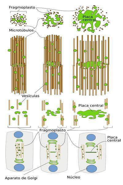
During the process of cell plate formation, the fragmoplast is formed. This is assembled from the remains of the mitotic spindle and is made up of a series of polar microtubules that apparently arise from the remains of the mitotic spindle apparatus and are organized in an antiparallel matrix..
These microtubules are aligned perpendicular to the plane of division with their “+” ends located at or near the site of cell division, and their negative ends face the two daughter nuclei..
The so-called "+" ends are the fast growing ends and are the place where the microtubules bond. Therefore, it is important to note that these “+” ends are immersed in an electrodense material located in the central zone..
In the later phase of anaphase, the slightly extended microtubules in the intermediate zone unite laterally in a cylindrical structure, the fragmoplast itself.
This structure subsequently shortens in length and expands laterally until it finally reaches the side wall. During this stage of fragmoplast expansion, a change occurs in the organization of microtubules..
Whereas the initial fragmoplast cylinder originates from pre-existing microtubules, new microtubules must be formed in the later stages of centrifugal growth..
Actin microfilaments are also an important cytoskeletal component of fragmoplasts. Their alignment, like that of microtubules, is perpendicular to the plane of the cell plate, with the “+” ends directed proximally.
Unlike microtubules, they are organized into two opposite sets that do not overlap or join directly. With the positive proximal ends, the actin microfilaments are also organized in a way that would facilitate the transport of vesicles to the plane of the plate..
The site where cell division will occur is established from a rearrangement of the microtubules that form the preprophase band, the mitotic spindle and the fragmoplast. When mitosis begins, the microtubules depolymerize and rearrange, forming the preprophase band around the nucleus..
Subsequently, the vesicles directed from the trans Golgi network (network of cellular structures and cisterns of the Golgi apparatus) towards the fragmoplast fuse and give rise to the cell plate. Then, the bipolar organization of the microtubules allows the directional transport of the vesicles to the site of cell division.
Finally, the microtubules, actin filaments from the fragmoplast, and the cell plate expand centrifugally toward the periphery of the cell as cytokinesis proceeds, where the cell plate then attaches to the cell wall of the stem cell to complete the process of cytokinesis.
Yet No Comments